
Article Figures & Data
Figures
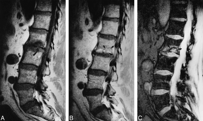
- fig 1.
Images of a 40-year-old man who experienced axial loading injury when thrown from a car and who presented with nonradiating lumbar pain and tenderness directly over the L3 and L5 vertebral bodies.
A, Sagittal midline short inversion time inversion recovery (1400/15/2) image, obtained during the initial MR examination, shows vertebral body edema and mild compression of the superior surfaces of the L3 and L5 vertebral bodies, but no SN is present at either level. Both levels are consistent with simple endplate fractures. Note the smooth superior surface of vertebral bodies L3 and L5, with what appear to be intact cortical margins (straight arrows). The inferior endplate of L5 may also be slightly compressed (curved arrow).
B, Sagittal gradient-echo (450/12/1) MR image, obtained 2 months later when the patient experienced some but not complete resolution of pain and tenderness, shows that a chronic SN formed at the superior endplate of L5 (curved arrow). Note resolution of marrow edema at L5 with persistent but improved edema of L3 (straight arrow). Further follow-up did not show SN formation at L3. This case illustrates that some endplate fractures may evolve into SNs and some may not.
- fig 2.
MR images obtained 6 days after injury of a 38-year-old man who experienced the sudden onset of pain and point tenderness referable to the L2 level immediately after experiencing axial-loading injury.
A, T1-weighted sagittal (600/15/2) image shows subtle concave compression of the caudal L2 endplate without obvious disk extension but with localized marrow edema. The margin of the SN is not well delineated because of the marrow edema, but a very small cortical defect representing an endplate fracture is present (arrow).
B, After the administration of contrast material, enhancement of the marrow edema peripheral to and within the SN defect can be seen. The margins of the SN are slightly more visible after the injection of the contrast material (arrows).
C, Gradient-echo (400/15/1) image best shows endplate invagination of disk material, establishing this abnormality as an acute SN. The margin of the SN can be most clearly appreciated as a linear area of decreased signal surrounding the extruded disk material (arrows) on the gradient-echo image. At 4-month follow-up, when the patient's pain had resolved, the edema had disappeared but the appearance of the SN on T2-weighted images was otherwise unchanged.
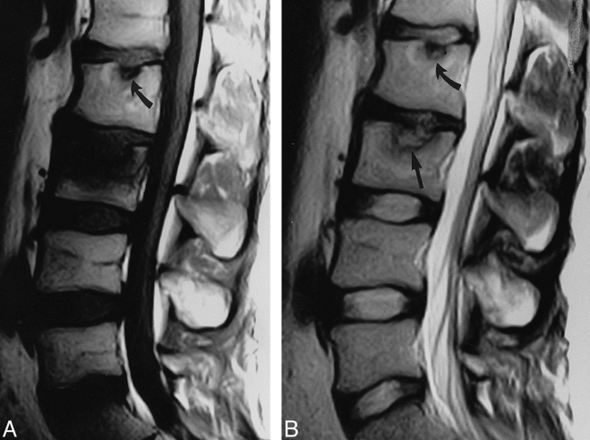
- fig 3.
Images of a 27-year-old man who presented with sudden onset of pain and tenderness centered at L3 after a snow ski jump injury involving pure axial loading.
A, Sagittal T1-weighted (600/15/2) image shows diffuse marrow edema of the cephalad two thirds of vertebral body L3, centered along the superior endplate. On the T1-weighted image, this looks like a simple endplate fracture. Note the nonacute SN and the narrowed disk space at L2 (arrow).
B, Sagittal fast spin-echo T2-weighted (4000/90/2) image, obtained as part of the same examination, shows the margins of the acute SN at L3 to better advantage (straight arrow). The L2 SN is unchanged (curved arrow).
- fig 4.
Images of a 45-year-old man who had experienced axial-loading injury 4 years before these images were obtained and who experienced the sudden onset of acute nonradiating low back pain with tenderness over L3.
A, Sagittal midline T1-weighted (700/15/2) image shows a small, nonacute SN at the inferior endplate of L3 with surrounding degenerative changes (arrow) and additional SNs without degenerative marrow changes at T12 to L2. The patient no longer experienced the pain and tenderness noted at the time of the original injury, and the original MR images are no longer in existence.
B, After the IV administration of contrast material, central enhancement within the asymptomatic SN was noted, probably in the granulation tissue that formed within the extruded fragment of disk (arrow). This illustrates one of the enhancement patterns that might be encountered with SNs. The other SNs from T12 to L2 do not enhance.


Tables

Summary of findings on initial MR examination in 14 patients with acute-onset low back pain after axial loading trauma


