Advertisement

Main menu

  • Home
  • Content
    • Current Issue
    • Accepted Manuscripts
    • Article Preview
    • Past Issue Archive
    • Video Articles
    • AJNR Case Collection
    • Case of the Week Archive
    • Case of the Month Archive
    • Classic Case Archive
  • Special Collections
    • AJNR Awards
    • Low-Field MRI
    • Alzheimer Disease
    • ASNR Foundation Special Collection
    • Photon-Counting CT
    • View All
  • Multimedia
    • AJNR Podcasts
    • AJNR SCANtastic
    • Trainee Corner
    • MRI Safety Corner
    • Imaging Protocols
  • For Authors
    • Submit a Manuscript
    • Submit a Video Article
    • Submit an eLetter to the Editor/Response
    • Manuscript Submission Guidelines
    • Statistical Tips
    • Fast Publishing of Accepted Manuscripts
    • Graphical Abstract Preparation
    • Imaging Protocol Submission
    • Author Policies
  • About Us
    • About AJNR
    • Editorial Board
    • Editorial Board Alumni
  • More
    • Become a Reviewer/Academy of Reviewers
    • Subscribers
    • Permissions
    • Alerts
    • Feedback
    • Advertisers
    • ASNR Home

User menu

  • Alerts
  • Log in

Search

  • Advanced search
American Journal of Neuroradiology

American Journal of Neuroradiology

ASHNR American Society of Functional Neuroradiology ASHNR American Society of Pediatric Neuroradiology ASSR
  • Alerts
  • Log in

Advanced Search

  • Home
  • Content
    • Current Issue
    • Accepted Manuscripts
    • Article Preview
    • Past Issue Archive
    • Video Articles
    • AJNR Case Collection
    • Case of the Week Archive
    • Case of the Month Archive
    • Classic Case Archive
  • Special Collections
    • AJNR Awards
    • Low-Field MRI
    • Alzheimer Disease
    • ASNR Foundation Special Collection
    • Photon-Counting CT
    • View All
  • Multimedia
    • AJNR Podcasts
    • AJNR SCANtastic
    • Trainee Corner
    • MRI Safety Corner
    • Imaging Protocols
  • For Authors
    • Submit a Manuscript
    • Submit a Video Article
    • Submit an eLetter to the Editor/Response
    • Manuscript Submission Guidelines
    • Statistical Tips
    • Fast Publishing of Accepted Manuscripts
    • Graphical Abstract Preparation
    • Imaging Protocol Submission
    • Author Policies
  • About Us
    • About AJNR
    • Editorial Board
    • Editorial Board Alumni
  • More
    • Become a Reviewer/Academy of Reviewers
    • Subscribers
    • Permissions
    • Alerts
    • Feedback
    • Advertisers
    • ASNR Home
  • Follow AJNR on Twitter
  • Visit AJNR on Facebook
  • Follow AJNR on Instagram
  • Join AJNR on LinkedIn
  • RSS Feeds

AJNR Awards, New Junior Editors, and more. Read the latest AJNR updates

Research ArticleBRAIN

Apoptosis in Leukoaraiosis

William R. Brown, Dixon M. Moody, Clara R. Thore and Venkata R. Challa
American Journal of Neuroradiology January 2000, 21 (1) 79-82;
William R. Brown
aFrom the Departments of Radiology (W.R.B., D.M.M., C.R.T.) and Pathology (W.R.B., V.R.C.) and the Program in Neuroscience (W.R.B., D.M.M.), Wake Forest University School of Medicine, Winston-Salem, NC 27157.
  • Find this author on Google Scholar
  • Find this author on PubMed
  • Search for this author on this site
Dixon M. Moody
aFrom the Departments of Radiology (W.R.B., D.M.M., C.R.T.) and Pathology (W.R.B., V.R.C.) and the Program in Neuroscience (W.R.B., D.M.M.), Wake Forest University School of Medicine, Winston-Salem, NC 27157.
  • Find this author on Google Scholar
  • Find this author on PubMed
  • Search for this author on this site
Clara R. Thore
aFrom the Departments of Radiology (W.R.B., D.M.M., C.R.T.) and Pathology (W.R.B., V.R.C.) and the Program in Neuroscience (W.R.B., D.M.M.), Wake Forest University School of Medicine, Winston-Salem, NC 27157.
  • Find this author on Google Scholar
  • Find this author on PubMed
  • Search for this author on this site
Venkata R. Challa
aFrom the Departments of Radiology (W.R.B., D.M.M., C.R.T.) and Pathology (W.R.B., V.R.C.) and the Program in Neuroscience (W.R.B., D.M.M.), Wake Forest University School of Medicine, Winston-Salem, NC 27157.
  • Find this author on Google Scholar
  • Find this author on PubMed
  • Search for this author on this site
  • Article
  • Figures & Data
  • Info & Metrics
  • Responses
  • References
  • PDF
Loading

Article Figures & Data

Figures

  • Tables
  • fig 1.
    • Download figure
    • Open in new tab
    • Download powerpoint
    fig 1.

    Spin-echo MR image (2600/20/1 [TR/TE/excitations]) of a 1.5-cm-thick coronal slice from the frontal region of the brain. Only the left hemisphere is shown. The open arrows indicate an area of leukoaraiosis (white matter hyperintensity) that corresponds to the area that was investigated for apoptosis (see fig 2). C, cortex of the sulcus identified in fig 2

  • fig 2.
    • Download figure
    • Open in new tab
    • Download powerpoint
    fig 2.

    Luxol fast blue–stained section from the brain slice imaged in figure 1. The outlined area corresponds to the area of leukoaraiosis in the MR image in figure 1. Reduced Luxol fast blue staining in this area indicates demyelination. Note the darker blue staining (indicating intact myelin) of the U fibers adjacent to the cortex (C) at the bottom of a sulcus. V, lateral ventricle.fig 3. Trichrome staining shows excessively thick collagen layers (green) in the walls of small veins and venules (arrows) in the area of demyelination, where these thick-walled veins were most numerous. They are usually more numerous near the angle of the lateral ventricle.fig 4. TUNEL staining in the area of demyelination shows positive cells (brown stain) in the wall of a blood vessel (arrowheads) and in the brain parenchyma (arrow). Inset A, The brown, TUNEL-stained cell in the parenchyma appears to be apoptotic histologically in that its blue-stained nucleus is condensed and split into two segments. Inset B, Another TUNEL-stained cell in the lesional white matter with nuclear fragmentation.fig 5. A moderate number of amyloid plaques are shown in the area of cortex that was counted. Inset, Double stained for β-amyloid (black) and interleukin-1 (red) (the latter stains activated microglia and macrophages)
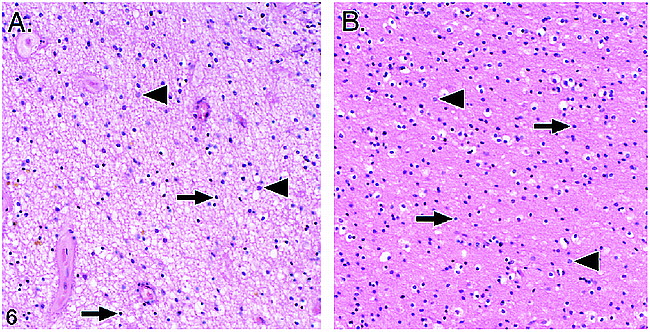
  • fig 6.
    • Download figure
    • Open in new tab
    • Download powerpoint
    fig 6.

    Comparison of the leukoaraiosis lesion (A) and an unaffected area in nearby white matter (B). Note that in the leukoaraiosis lesion, oligodendrocytes (arrows) appear to be preferentially less numerous than astrocytes, which have nuclei that are slightly larger and less densely stained (arrowheads) (hematoxylin and eosin)

Tables

  • Figures
  • Table1

    TABLE: Apoptotic cell counts/cm2 in three areas in five sections

PreviousNext
Back to top

In this issue

American Journal of Neuroradiology
Vol. 21, Issue 1
1 Jan 2000
  • Table of Contents
  • Index by author
Advertisement
Print
Download PDF
Email Article

Thank you for your interest in spreading the word on American Journal of Neuroradiology.

NOTE: We only request your email address so that the person you are recommending the page to knows that you wanted them to see it, and that it is not junk mail. We do not capture any email address.

Enter multiple addresses on separate lines or separate them with commas.
Apoptosis in Leukoaraiosis
(Your Name) has sent you a message from American Journal of Neuroradiology
(Your Name) thought you would like to see the American Journal of Neuroradiology web site.
CAPTCHA
This question is for testing whether or not you are a human visitor and to prevent automated spam submissions.
Cite this article
William R. Brown, Dixon M. Moody, Clara R. Thore, Venkata R. Challa
Apoptosis in Leukoaraiosis
American Journal of Neuroradiology Jan 2000, 21 (1) 79-82;

Citation Manager Formats

  • BibTeX
  • Bookends
  • EasyBib
  • EndNote (tagged)
  • EndNote 8 (xml)
  • Medlars
  • Mendeley
  • Papers
  • RefWorks Tagged
  • Ref Manager
  • RIS
  • Zotero
0 Responses
Respond to this article
Share
Bookmark this article
Apoptosis in Leukoaraiosis
William R. Brown, Dixon M. Moody, Clara R. Thore, Venkata R. Challa
American Journal of Neuroradiology Jan 2000, 21 (1) 79-82;
del.icio.us logo Twitter logo Facebook logo Mendeley logo
  • Tweet Widget
  • Facebook Like
  • Google Plus One

Jump to section

  • Article
    • Abstract
    • Case Report
    • Discussion
    • Acknowledgments
    • Footnotes
    • References
  • Figures & Data
  • Info & Metrics
  • Responses
  • References
  • PDF

Related Articles

  • No related articles found.
  • PubMed
  • Google Scholar

Cited By...

  • Longitudinal evidence for a mutually reinforcing relationship between white matter hyperintensities and cortical thickness in cognitively unimpaired older adults
  • Automated White Matter Total Lesion Volume Segmentation in Diabetes
  • Understanding White Matter Disease: Imaging-Pathological Correlations in Vascular Cognitive Impairment
  • Leukoaraiosis: The brain under pressure: Target for treatment?
  • White Matter Lesions and Glial Activation in a Novel Mouse Model of Chronic Cerebral Hypoperfusion
  • Patients with vascular dementia due to microvascular pathology have significant hippocampal neuronal loss
  • Cerebral Autosomal Dominant Arteriopathy with Subcortical Infarcts and Leukoencephalopathy: Decrease in Regional Cerebral Blood Volume in Hyperintense Subcortical Lesions Inversely Correlates with Disability and Cognitive Performance
  • Crossref
  • Google Scholar

This article has not yet been cited by articles in journals that are participating in Crossref Cited-by Linking.

More in this TOC Section

  • Progression of Microstructural Damage in Spinocerebellar Ataxia Type 2: A Longitudinal DTI Study
  • SWI or T2*: Which MRI Sequence to Use in the Detection of Cerebral Microbleeds? The Karolinska Imaging Dementia Study
  • Statin Therapy Does Not Affect the Radiographic and Clinical Profile of Patients with TIA and Minor Stroke
Show more Brain

Similar Articles

Advertisement

Indexed Content

  • Current Issue
  • Accepted Manuscripts
  • Article Preview
  • Past Issues
  • Editorials
  • Editor's Choice
  • Fellows' Journal Club
  • Letters to the Editor
  • Video Articles

Cases

  • Case Collection
  • Archive - Case of the Week
  • Archive - Case of the Month
  • Archive - Classic Case

More from AJNR

  • Trainee Corner
  • Imaging Protocols
  • MRI Safety Corner
  • Book Reviews

Multimedia

  • AJNR Podcasts
  • AJNR Scantastics

Resources

  • Turnaround Time
  • Submit a Manuscript
  • Submit a Video Article
  • Submit an eLetter to the Editor/Response
  • Manuscript Submission Guidelines
  • Statistical Tips
  • Fast Publishing of Accepted Manuscripts
  • Graphical Abstract Preparation
  • Imaging Protocol Submission
  • Evidence-Based Medicine Level Guide
  • Publishing Checklists
  • Author Policies
  • Become a Reviewer/Academy of Reviewers
  • News and Updates

About Us

  • About AJNR
  • Editorial Board
  • Editorial Board Alumni
  • Alerts
  • Permissions
  • Not an AJNR Subscriber? Join Now
  • Advertise with Us
  • Librarian Resources
  • Feedback
  • Terms and Conditions
  • AJNR Editorial Board Alumni

American Society of Neuroradiology

  • Not an ASNR Member? Join Now

© 2025 by the American Society of Neuroradiology All rights, including for text and data mining, AI training, and similar technologies, are reserved.
Print ISSN: 0195-6108 Online ISSN: 1936-959X

Powered by HighWire