
Article Figures & Data
Figures
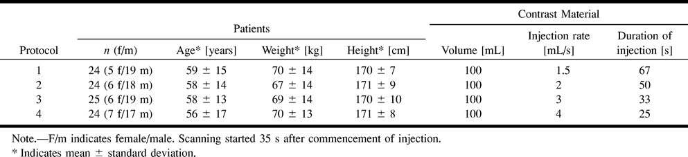
- fig 1.
Times of contrast material injection versus scan time in four protocols with different contrast material injection rates
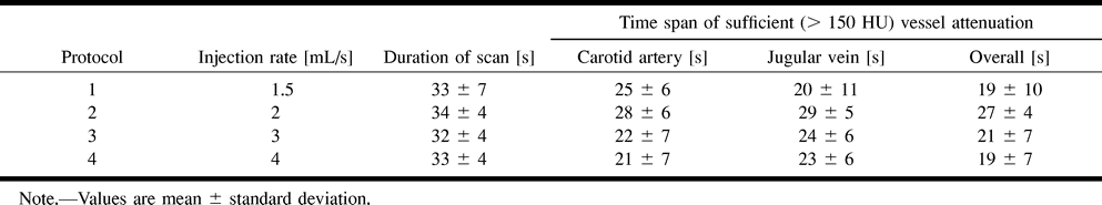
- fig 2.
Mean time-density curves.
A and B, Curves of the carotid artery and the jugular vein with four protocols of different contrast material injection rates.
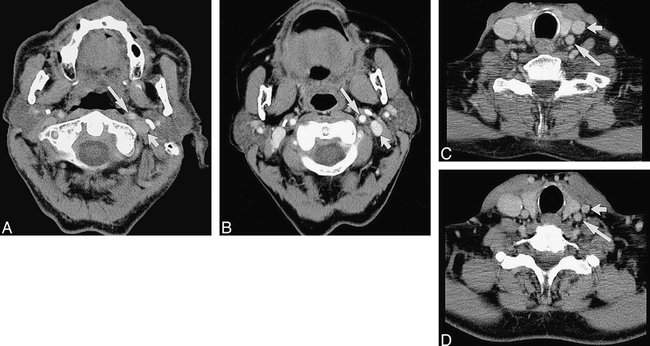
- fig 3.
Representative CT images using four different contrast material injection rates.
A, The 1.5 mL/s protocol results in moderate attenuation of the carotid artery (long arrow) but insufficient attenuation of the jugular vein (short arrow).
B, The longest times of sufficient vessel attenuation are achieved with the 2 mL/s protocol.
C and D, With the 3 mL/s and 4 mL/s protocols the attenuation of the carotid artery and the jugular vein show a significant decrease towards the end of the scan sequence.


