Advertisement

Main menu

  • Home
  • Content
    • Current Issue
    • Accepted Manuscripts
    • Article Preview
    • Past Issue Archive
    • Video Articles
    • AJNR Case Collection
    • Case of the Week Archive
    • Case of the Month Archive
    • Classic Case Archive
  • Special Collections
    • AJNR Awards
    • Low-Field MRI
    • Alzheimer Disease
    • ASNR Foundation Special Collection
    • Photon-Counting CT
    • View All
  • Multimedia
    • AJNR Podcasts
    • AJNR SCANtastic
    • Trainee Corner
    • MRI Safety Corner
    • Imaging Protocols
  • For Authors
    • Submit a Manuscript
    • Submit a Video Article
    • Submit an eLetter to the Editor/Response
    • Manuscript Submission Guidelines
    • Statistical Tips
    • Fast Publishing of Accepted Manuscripts
    • Graphical Abstract Preparation
    • Imaging Protocol Submission
    • Author Policies
  • About Us
    • About AJNR
    • Editorial Board
    • Editorial Board Alumni
  • More
    • Become a Reviewer/Academy of Reviewers
    • Subscribers
    • Permissions
    • Alerts
    • Feedback
    • Advertisers
    • ASNR Home

User menu

  • Alerts
  • Log in

Search

  • Advanced search
American Journal of Neuroradiology

American Journal of Neuroradiology

ASHNR American Society of Functional Neuroradiology ASHNR American Society of Pediatric Neuroradiology ASSR
  • Alerts
  • Log in

Advanced Search

  • Home
  • Content
    • Current Issue
    • Accepted Manuscripts
    • Article Preview
    • Past Issue Archive
    • Video Articles
    • AJNR Case Collection
    • Case of the Week Archive
    • Case of the Month Archive
    • Classic Case Archive
  • Special Collections
    • AJNR Awards
    • Low-Field MRI
    • Alzheimer Disease
    • ASNR Foundation Special Collection
    • Photon-Counting CT
    • View All
  • Multimedia
    • AJNR Podcasts
    • AJNR SCANtastic
    • Trainee Corner
    • MRI Safety Corner
    • Imaging Protocols
  • For Authors
    • Submit a Manuscript
    • Submit a Video Article
    • Submit an eLetter to the Editor/Response
    • Manuscript Submission Guidelines
    • Statistical Tips
    • Fast Publishing of Accepted Manuscripts
    • Graphical Abstract Preparation
    • Imaging Protocol Submission
    • Author Policies
  • About Us
    • About AJNR
    • Editorial Board
    • Editorial Board Alumni
  • More
    • Become a Reviewer/Academy of Reviewers
    • Subscribers
    • Permissions
    • Alerts
    • Feedback
    • Advertisers
    • ASNR Home
  • Follow AJNR on Twitter
  • Visit AJNR on Facebook
  • Follow AJNR on Instagram
  • Join AJNR on LinkedIn
  • RSS Feeds

AJNR Awards, New Junior Editors, and more. Read the latest AJNR updates

Research ArticlePEDIATRICS

MR Line-Scan Diffusion-Weighted Imaging of Term Neonates with Perinatal Brain Ischemia

Richard L. Robertson, Liat Ben-Sira, Patrick D. Barnes, Robert V. Mulkern, Caroline D. Robson, Stephan E. Maier, Michael J. Rivkin and Adre J. du Plessis
American Journal of Neuroradiology October 1999, 20 (9) 1658-1670;
Richard L. Robertson
  • Find this author on Google Scholar
  • Find this author on PubMed
  • Search for this author on this site
Liat Ben-Sira
  • Find this author on Google Scholar
  • Find this author on PubMed
  • Search for this author on this site
Patrick D. Barnes
  • Find this author on Google Scholar
  • Find this author on PubMed
  • Search for this author on this site
Robert V. Mulkern
  • Find this author on Google Scholar
  • Find this author on PubMed
  • Search for this author on this site
Caroline D. Robson
  • Find this author on Google Scholar
  • Find this author on PubMed
  • Search for this author on this site
Stephan E. Maier
  • Find this author on Google Scholar
  • Find this author on PubMed
  • Search for this author on this site
Michael J. Rivkin
  • Find this author on Google Scholar
  • Find this author on PubMed
  • Search for this author on this site
Adre J. du Plessis
  • Find this author on Google Scholar
  • Find this author on PubMed
  • Search for this author on this site
  • Article
  • Figures & Data
  • Info & Metrics
  • Responses
  • References
  • PDF
Loading

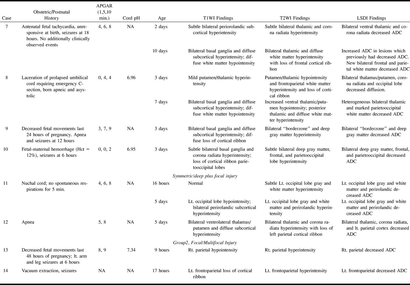
Article Figures & Data

Figures

  • Tables
  • fig 1.
    • Download figure
    • Open in new tab
    • Download powerpoint
    fig 1.

    Patient 1. Neonate of estimated 38-week gestational age with placental abruption and bradycardia.

    A, Axial T2-weighted fast spin-echo image (3200/85/1), with an echo train length of 8, obtained at 13 hours of life shows no abnormality.

    B, Trace LSDI image (1520/62.5/1), with a b max of 750 seconds/mm2, obtained at 13 hours of life shows no abnormality.

    C, Corresponding ADC map.

    D, Axial T2-weighted fast spin-echo image obtained at 5 days of life shows very subtle hyperintensity in the posterior putamen bilaterally (arrows).

    E, Trace LSDI image obtained at 5 days of life shows decreased diffusion in corresponding areas (arrows).

    F, Corresponding ADC map.

    G, T1-weighted axial spin-echo image (600/20/2) obtained at 6 weeks of life shows hyperintensity within the posterior putamen and ventrolateral thalamus bilaterally (arrows).
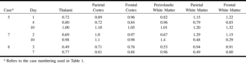
  • fig 2.
    • Download figure
    • Open in new tab
    • Download powerpoint
    fig 2.

    Patient 8. Neonate of estimated 39-week gestational age with a lacerated, prolapsed umbilical cord.

    A, Axial T2-weighted fast spin-echo image (3200/85/1), with an echo train length of 8, obtained at 3 days of life at the level of the basal ganglia shows T2 shortening (hypointensity) in the posterior putamen and ventrolateral thalamus bilaterally (compare with fig 1A). The cortical ribbon is indistinct.

    B, ADC map (1520/62.5/1), with a b max of 750 s/mm2, obtained at 3 days of life at a corresponding level shows decreased diffusion (hypointensity) in the ventrolateral thalami (arrows) and, to a lesser extent, in the occipital lobe gray matter (arrowheads). The hyperintensity of the frontal lobe white matter is due to the high diffusion that is normally present in the white matter of neonates.

    C, Axial T2-weighted image obtained at 3 days of life above the lateral ventricles shows loss of the cortical ribbon in the frontal and parietal lobes and mild white matter hyperintensity.

    D, ADC map obtained at 3 days of life at the same level shows pathologically decreased diffusion in the left perirolandic white matter (arrows). Decreased diffusion was apparent in the right perirolandic white matter at other levels (not shown). Note that the white matter of the centrum semiovale apart from the perirolandic white matter does not have decreased diffusion.

    E, Axial T2-weighted image obtained at 7 days of life shows marked hypointensity in the posterior putamen/ventrolateral thalami, posterior thalamic hyperintensity, and diffuse white matter hyperintensity.

    F, ADC map obtained at 7 days of life at a corresponding location shows heterogeneously decreased ADCs (hypointensity) within the thalami and markedly decreased ADCs throughout the occipital lobe white matter.

    G, Axial T2-weighted image obtained at 7 days of life shows marked T2 prolongation within the white matter.

    H, ADC map obtained at 7 days of life shows decreased ADCs within the white matter.
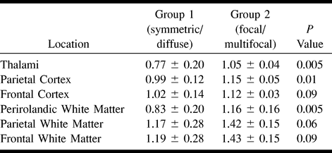
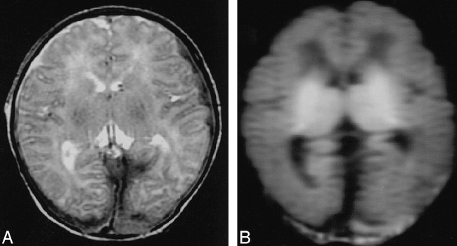
  • fig 3.
    • Download figure
    • Open in new tab
    • Download powerpoint
    fig 3.

    Patient 6. Neonate of estimated 41-week gestational age with placental abruption, no spontaneous respirations, and seizures. Life support was withdrawn at 23 hours of life.

    A, Axial T2-weighted fast spin-echo image (3200/85/1), with an echo train length of 8, obtained at 18 hours of life shows no brain abnormality. Small bilateral occipital subdural hematomas are present.

    B, Trace LSDI image (1520/62.5/1), with a b max of 750 seconds/mm2, shows decreased diffusion (hyperintensity) bilaterally in the basal ganglia and thalami. Note that the frontal and parietal lobe cortex and white matter appear to have normal diffusion. Pathologic analysis revealed diffuse infarction was present throughout the brain.
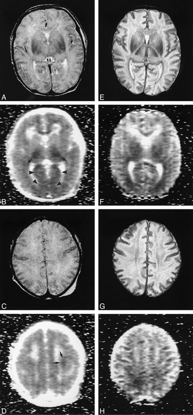
  • fig 4.
    • Download figure
    • Open in new tab
    • Download powerpoint
    fig 4.

    Patient 13. Neonate of estimated 38-week gestational age with decreased fetal movements during the last 48 hours of pregnancy and left arm and leg seizures at 6 hours of life.

    A, Axial T2-weighted fast spin-echo image (3200/85/1), with an echo train length of 8, obtained at 9 hours of life shows focal loss of the right parietal cortical ribbon.

    B, Trace LSDI image (1520/62.5/1), with a b max of 750 seconds/mm2, shows markedly decreased diffusion (hyperintensity) in a corresponding location.

    C, ADC map shows markedly decreased diffusion (hypointensity) within the lesion.

Tables

  • Figures
  • TABLE 1:
    • View popup
    • Download powerpoint
    TABLE 1:

    Clinical data and MR findings of 19 neonates with suspected perinatal brain ischemia

  • TABLE 1:
    • View popup
    • Download powerpoint
    TABLE 1:

    Continued

  • TABLE 1:
    • View popup
    • Download powerpoint
    TABLE 1:

    Continued

  • TABLE 2:
    • View popup
    • Download powerpoint
    TABLE 2:

    ADC measurements (μm2/ms) in three neonates with diffuse injury

  • TABLE 3:
    • View popup
    • Download powerpoint
    TABLE 3:

    Mean and standard deviation of ADC measurements (μm2/ms) in neonates imaged during the first 72 hours with symmetric/diffuse injury (Group 1) versus focal/multifocal injury (Group 2)

PreviousNext
Back to top

In this issue

American Journal of Neuroradiology
Vol. 20, Issue 9
1 Oct 1999
  • Table of Contents
  • Index by author
Advertisement
Print
Download PDF
Email Article

Thank you for your interest in spreading the word on American Journal of Neuroradiology.

NOTE: We only request your email address so that the person you are recommending the page to knows that you wanted them to see it, and that it is not junk mail. We do not capture any email address.

Enter multiple addresses on separate lines or separate them with commas.
MR Line-Scan Diffusion-Weighted Imaging of Term Neonates with Perinatal Brain Ischemia
(Your Name) has sent you a message from American Journal of Neuroradiology
(Your Name) thought you would like to see the American Journal of Neuroradiology web site.
CAPTCHA
This question is for testing whether or not you are a human visitor and to prevent automated spam submissions.
Cite this article
Richard L. Robertson, Liat Ben-Sira, Patrick D. Barnes, Robert V. Mulkern, Caroline D. Robson, Stephan E. Maier, Michael J. Rivkin, Adre J. du Plessis
MR Line-Scan Diffusion-Weighted Imaging of Term Neonates with Perinatal Brain Ischemia
American Journal of Neuroradiology Oct 1999, 20 (9) 1658-1670;

Citation Manager Formats

  • BibTeX
  • Bookends
  • EasyBib
  • EndNote (tagged)
  • EndNote 8 (xml)
  • Medlars
  • Mendeley
  • Papers
  • RefWorks Tagged
  • Ref Manager
  • RIS
  • Zotero
0 Responses
Respond to this article
Share
Bookmark this article
MR Line-Scan Diffusion-Weighted Imaging of Term Neonates with Perinatal Brain Ischemia
Richard L. Robertson, Liat Ben-Sira, Patrick D. Barnes, Robert V. Mulkern, Caroline D. Robson, Stephan E. Maier, Michael J. Rivkin, Adre J. du Plessis
American Journal of Neuroradiology Oct 1999, 20 (9) 1658-1670;
del.icio.us logo Twitter logo Facebook logo Mendeley logo
  • Tweet Widget
  • Facebook Like
  • Google Plus One

Jump to section

  • Article
    • Abstract
    • Methods
    • Results
    • Discussion
    • Conclusion
    • Footnotes
    • References
  • Figures & Data
  • Info & Metrics
  • Responses
  • References
  • PDF

Related Articles

  • No related articles found.
  • PubMed
  • Google Scholar

Cited By...

  • Evolution of Unilateral Perinatal Arterial Ischemic Stroke on Conventional and Diffusion-Weighted MR Imaging
  • Neonatal Neurologic Consultations: Integration With Maternal-fetal Medicine and Long-term Outcome
  • Early diffusion weighted imaging and expression of heat shock protein 70 in newborn pigs with hypoxic ischaemic encephalopathy
  • Recovery of amplitude integrated electroencephalographic background patterns within 24 hours of perinatal asphyxia
  • A prospective, longitudinal diffusion tensor imaging study of brain injury in newborns
  • Magnetic resonance imaging of the infant brain: anatomical characteristics and clinical significance of punctate lesions
  • Proton Spectroscopy and Diffusion Imaging on the First Day of Life after Perinatal Asphyxia: Preliminary Report
  • Magnetic Resonance Imaging of the Fetal and Neonatal Central Nervous System
  • Neonatal Hypoxic-ischemic Encephalopathy: Detection with Diffusion-weighted MR Imaging
  • Crossref
  • Google Scholar

This article has not yet been cited by articles in journals that are participating in Crossref Cited-by Linking.

More in this TOC Section

  • Early Ultrasonic Monitoring of Brain Growth and Later Neurodevelopmental Outcome in Very Preterm Infants
  • Diagnostic Value of Sylvian Fissure Hyperechogenicity in Fetal SAH
  • Feasibility and Added Value of Fetal DTI Tractography in the Evaluation of an Isolated Short Corpus Callosum: Preliminary Results
Show more Pediatrics

Similar Articles

Advertisement

Indexed Content

  • Current Issue
  • Accepted Manuscripts
  • Article Preview
  • Past Issues
  • Editorials
  • Editor's Choice
  • Fellows' Journal Club
  • Letters to the Editor
  • Video Articles

Cases

  • Case Collection
  • Archive - Case of the Week
  • Archive - Case of the Month
  • Archive - Classic Case

More from AJNR

  • Trainee Corner
  • Imaging Protocols
  • MRI Safety Corner
  • Book Reviews

Multimedia

  • AJNR Podcasts
  • AJNR Scantastics

Resources

  • Turnaround Time
  • Submit a Manuscript
  • Submit a Video Article
  • Submit an eLetter to the Editor/Response
  • Manuscript Submission Guidelines
  • Statistical Tips
  • Fast Publishing of Accepted Manuscripts
  • Graphical Abstract Preparation
  • Imaging Protocol Submission
  • Evidence-Based Medicine Level Guide
  • Publishing Checklists
  • Author Policies
  • Become a Reviewer/Academy of Reviewers
  • News and Updates

About Us

  • About AJNR
  • Editorial Board
  • Editorial Board Alumni
  • Alerts
  • Permissions
  • Not an AJNR Subscriber? Join Now
  • Advertise with Us
  • Librarian Resources
  • Feedback
  • Terms and Conditions
  • AJNR Editorial Board Alumni

American Society of Neuroradiology

  • Not an ASNR Member? Join Now

© 2025 by the American Society of Neuroradiology All rights, including for text and data mining, AI training, and similar technologies, are reserved.
Print ISSN: 0195-6108 Online ISSN: 1936-959X

Powered by HighWire