
Article Figures & Data
Figures
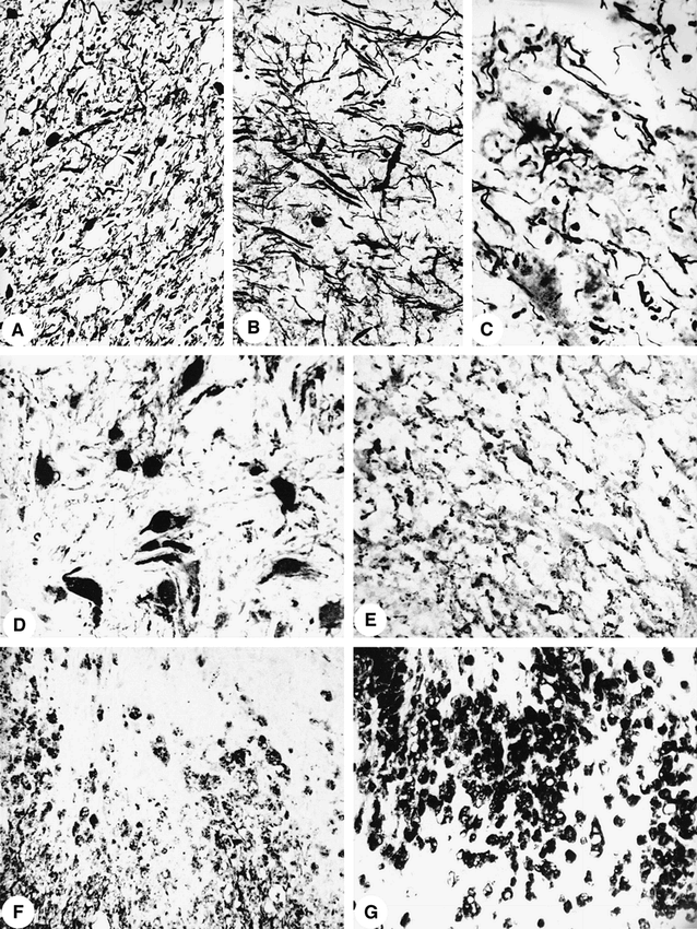
- fig 1.
Bielschowsky's silver impregnation shows axonal density in the normal periplaque white matter (patient 1) (A), a reduction by 45% (patient 3) (B), and a reduction by 75% (patient 1) (C) within the plaques (magnification for all, ×166). Prominent protoplasmic gliosis with large astrocytes (D) is seen by immunocytochemistry for GFAP (patient 1). Numerous GFAP-positive cell processes (E) indicate fibrillary gliosis (patient 3). Immunocytochemistry for MOG (F) shows an early active demyelinating lesion (patient 2). Macrophages carry MOG-positive degradation products in their cytoplasm. Dense macrophage infiltrate (G) is seen in the same lesion as shown in F (immunocytochemistry for Ki-MlP)
- fig 2.
T1-weighted gradient-echo MR images (3D fast low-angle shot, 4-mm partitions, 15/6 TR/TE, 20° flip angle) of patient 1 indicating VOIs selected for MRS.
A, Transverse section with VOIs centered at the left parieto-occipital lesion (20 × 20 × 20 mm3) and in a contralateral control region (16 × 30 × 16 mm3).
B, Sagittal section with a smaller VOI (16 × 16 × 16 mm3) encompassing the same lesion (note small defect in the skull and corresponding biopsy canal),
C, Coronal section depicting an ipsilateral VOI (20 × 20 × 20 mm3) in left frontoparietal cortex unsuspicious at MR imaging.
- fig 3.
Localized proton MR spectra (stimulated-echo acquisition mode, 3000/20/30 [TR/TE/TM]) of patient 1 from locations indicated in figure 2. Left occipitoparietal lesion (top) image using a 4-mL VOI and (second row) 8-mL VOI, an ipsilateral 8-mL VOI in unsuspicious left frontoparietal cortex (third row), and contralateral control (bottom). Major resonances are due to N-acetylaspartate (NAA), creatine and phosphocreatine (Cr), choline-containing compounds (Cho), myo-inositol (Ins), and lactate (Lac). Spectra are normalized for comparison


Tables

- Table 2:
Lesion histopathology and corresponding proton MRS-detected metabolic alterations. Absolute metabolite levels (mM;shL VOI) derived from proton MR spectra (STEAM, TR;shTE;shTM = 3000;sh20;sh30 ms) of biopsied lesions of patients ;ns1 to ;ns3


In this issue


Jump to section
Related Articles
Cited By...
- In vivo assessment of astrocyte reactivity in patients with progressive supranuclear palsy
- Linking cortical lesions to metabolic changes in multiple sclerosis using 7T proton MR spectroscopy
- The proton-activated receptor TDAG8 is upregulated in oligodendrocytes during maturation and under acidic conditions
- Alterations of Brain Metabolites in Adults With HIV: A Systematic Meta-analysis of Magnetic Resonance Spectroscopy Studies
- BRAIN MR SPECTROSCOPIC FINDINGS IN THREE CONSECUTIVE COVID-19 PATIENTS: PRELIMINARY OBSERVATIONS
- Brain MR Spectroscopic Findings in 3 Consecutive Patients with COVID-19: Preliminary Observations
- NAA is a Marker of Disability in Secondary-Progressive MS: A Proton MR Spectroscopic Imaging Study
- Regional Myo-Inositol, Creatine, and Choline Levels Are Higher at Older Age and Scale Negatively with Visuospatial Working Memory: A Cross-Sectional Proton MR Spectroscopy Study at 7 Tesla on Normal Cognitive Ageing
- Magnetic Resonance Imaging in Multiple Sclerosis
- Osmoregulatory inositol transporter SMIT1 modulates electrical activity by adjusting PI(4,5)P2 levels
- Early Alzheimer's Disease Neuropathology Detected by Proton MR Spectroscopy
- Utility of Proton MR Spectroscopy for Differentiating Typical and Atypical Primary Central Nervous System Lymphomas from Tumefactive Demyelinating Lesions
- Serial proton MR spectroscopy of gray and white matter in relapsing-remitting MS
- Metabolic profile of PML lesions in patients with and without IRIS: An observational study
- Correlating Quantitative MR Imaging with Histopathology in X-Linked Adrenoleukodystrophy
- Intracranial dural arteriovenous fistula presenting as an enhancing lesion of the medulla
- MRS in presymptomatic MAPT mutation carriers: A potential biomarker for tau-mediated pathology
- Diffuse White Matter Damage Is Absent in Neuromyelitis Optica
- MR spectroscopy indicates diffuse multiple sclerosis activity during remission
- Role of MRI in the differentiation of ADEM from MS in children
- Proton MR Spectroscopy Improves Discrimination between Tumor and Pseudotumoral Lesion in Solid Brain Masses
- 1H Magnetic resonance spectroscopy in dementia
- Guidelines for using proton MR spectroscopy in multicenter clinical MS studies
- Frontal brain lobe impairment in obstructive sleep apnoea: a proton MR spectroscopy study
- 1H MR spectroscopy in common dementias
- Five new cases of a recently described leukoencephalopathy with high brain lactate
- Inflammatory Demyelinating Disease Mimicking Malignant Glioma
- Brain N-acetylaspartate is elevated in Pelizaeus-Merzbacher disease with PLP1 duplication
- Magnetic resonance spectroscopy in AD
- Altered white and gray matter metabolism in CADASIL: A proton MR spectroscopy and 1H-MRSI study
- The Choline/Creatine Ratio in Five Benign Neoplasms: Comparison with Squamous Cell Carcinoma by Use of in Vitro MR Spectroscopy
- Serial Proton MR Spectroscopy of Contrast-enhancing Multiple Sclerosis Plaques: Absolute Metabolic Values over 2 Years during a Clinical Pharmacological Study