
Article Figures & Data
Figures
- fig 1.
A–C, Quantitative and qualitative analysis among the six sequences for CNR (A), lesion detection (B), and conspicuity of internal architecture (C). GRE indicates gradient-echo sequence; GRE-EPI, GRE-type echo-planar imaging; SE-EPI, spin-echo–type echo-planar imaging; TSE, turbo spin-echo sequence; HASTE, half-Fourier acquisition single-shot turbo spin-echo sequence; s-HASTE, segmented-HASTE sequence
- fig 2.
Intraventricular anaplastic astrocytoma in a 19-year-old woman.
A and B, The GRE (A) and GRE-EPI (B) images clearly show the intratumoral hemorrhage; however, the size of the hypointense area is larger on the GRE-EPI image than on the GRE image. The linear hemorrhage along the right ventricular wall (arrows) is seen on the GRE image only.
C and D, Contrast between the hemorrhage and the white matter is poorer on the SE-EPI (C) and TSE (D) images than on the GRE and GRE-EPI images.
E and F, Hemorrhage is poorly seen on the s-HASTE (E) and HASTE (F) images.
- fig 3.
Anaplastic astrocytoma after surgery and radio/chemotherapy in a 32-year-old woman.
A and B, Multiple small hemorrhagic lesions (large arrows) are detected on the GRE (A) and GRE-EPI (B) images.The GRE image is more sensitive to the cortical and subcortical lesions (arrowheads) than the GRE-EPI image, which is distorted by artifacts from frontal sinuses and calvaria. The GRE-EPI image is more sensitive to the white matter lesions than is the GRE image (small arrows).
C and D, Fewer lesions are seen on the SE-EPI (C) and TSE (D) images than on the GRE and GRE-EPI images (arrows and arrowheads).
E and F, The s-HASTE (E) and HASTE (F) images do not show the small hemorrhagic lesions (arrows and arrowheads). Note the interface low intensity between CSF and cerebral parenchyma on the s-HASTE image, which should not be mistaken for hemosiderosis (curved arrow).
G and H, Lower sections around the skull base. Susceptibility artifacts (arrows) from the skull base obscure the lesions at the infratentorial and skull-base level on the GRE-EPI image (H) and less so on the GRE image (G).


Tables

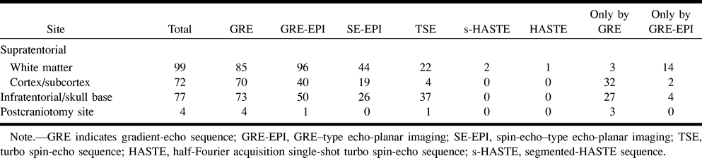
- TABLE 2:
Number of small lesions ([cf3]<[cf3]5 mm) detected by the six sequences

In this issue


Jump to section
Related Articles
Cited By...
- Understanding Lesion Progression in a Chronic Model of Cerebral Cavernous Malformations through Combined MRI and Histology
- Susceptibility-weighted MRI in mild traumatic brain injury
- Radiation Risk Due to Shunted Hydrocephalus and the Role of MR Imaging-Safe Programmable Valves
- A longitudinal MRI study of traumatic axonal injury in patients with moderate and severe traumatic brain injury
- Susceptibility-Weighted Imaging in Patients with Pyogenic Brain Abscesses at 1.5T: Characteristics of the Abscess Capsule
- Improved Delineation of Ventricular Shunt Catheters Using Fast Steady-State Gradient Recalled-Echo Sequences in a Rapid Brain MR Imaging Protocol in Nonsedated Pediatric Patients
- Susceptibility-Weighted Imaging in the Diagnosis of Early Basal Ganglia Germinoma
- Cerebral microbleeds in the population based AGES-Reykjavik study: prevalence and location
- Cerebral Microhemorrhages Predict New Disabling or Fatal Strokes in Patients With Acute Ischemic Stroke or Transient Ischemic Attack
- Contribution of Susceptibility-Weighted Imaging to Acute Stroke Assessment
- Detection of Intracranial Hemorrhage: Comparison between Gradient-echo Images and b0 Images Obtained from Diffusion-weighted Echo-planar Sequences
- Assessment of Lacunar Hemorrhage Associated With Hypertensive Stroke by Echo-Planar Gradient-Echo T2*-Weighted MRI