Advertisement

Main menu

  • Home
  • Content
    • Current Issue
    • Accepted Manuscripts
    • Article Preview
    • Past Issue Archive
    • Video Articles
    • AJNR Case Collection
    • Case of the Week Archive
    • Case of the Month Archive
    • Classic Case Archive
  • Special Collections
    • AJNR Awards
    • Low-Field MRI
    • Alzheimer Disease
    • ASNR Foundation Special Collection
    • Photon-Counting CT
    • View All
  • Multimedia
    • AJNR Podcasts
    • AJNR SCANtastic
    • Trainee Corner
    • MRI Safety Corner
    • Imaging Protocols
  • For Authors
    • Submit a Manuscript
    • Submit a Video Article
    • Submit an eLetter to the Editor/Response
    • Manuscript Submission Guidelines
    • Statistical Tips
    • Fast Publishing of Accepted Manuscripts
    • Graphical Abstract Preparation
    • Imaging Protocol Submission
    • Author Policies
  • About Us
    • About AJNR
    • Editorial Board
    • Editorial Board Alumni
  • More
    • Become a Reviewer/Academy of Reviewers
    • Subscribers
    • Permissions
    • Alerts
    • Feedback
    • Advertisers
    • ASNR Home

User menu

  • Alerts
  • Log in

Search

  • Advanced search
American Journal of Neuroradiology

American Journal of Neuroradiology

ASHNR American Society of Functional Neuroradiology ASHNR American Society of Pediatric Neuroradiology ASSR
  • Alerts
  • Log in

Advanced Search

  • Home
  • Content
    • Current Issue
    • Accepted Manuscripts
    • Article Preview
    • Past Issue Archive
    • Video Articles
    • AJNR Case Collection
    • Case of the Week Archive
    • Case of the Month Archive
    • Classic Case Archive
  • Special Collections
    • AJNR Awards
    • Low-Field MRI
    • Alzheimer Disease
    • ASNR Foundation Special Collection
    • Photon-Counting CT
    • View All
  • Multimedia
    • AJNR Podcasts
    • AJNR SCANtastic
    • Trainee Corner
    • MRI Safety Corner
    • Imaging Protocols
  • For Authors
    • Submit a Manuscript
    • Submit a Video Article
    • Submit an eLetter to the Editor/Response
    • Manuscript Submission Guidelines
    • Statistical Tips
    • Fast Publishing of Accepted Manuscripts
    • Graphical Abstract Preparation
    • Imaging Protocol Submission
    • Author Policies
  • About Us
    • About AJNR
    • Editorial Board
    • Editorial Board Alumni
  • More
    • Become a Reviewer/Academy of Reviewers
    • Subscribers
    • Permissions
    • Alerts
    • Feedback
    • Advertisers
    • ASNR Home
  • Follow AJNR on Twitter
  • Visit AJNR on Facebook
  • Follow AJNR on Instagram
  • Join AJNR on LinkedIn
  • RSS Feeds

AJNR Awards, New Junior Editors, and more. Read the latest AJNR updates

Research ArticleBrain

Evaluation of the Substantia Nigra in Patients with Parkinsonian Syndrome Accomplished Using Multishot Diffusion-Weighted MR Imaging

Michito Adachi, Takaaki Hosoya, Tamami Haku, Koichi Yamaguchi and Toru Kawanami
American Journal of Neuroradiology September 1999, 20 (8) 1500-1506;
Michito Adachi
  • Find this author on Google Scholar
  • Find this author on PubMed
  • Search for this author on this site
Takaaki Hosoya
  • Find this author on Google Scholar
  • Find this author on PubMed
  • Search for this author on this site
Tamami Haku
  • Find this author on Google Scholar
  • Find this author on PubMed
  • Search for this author on this site
Koichi Yamaguchi
  • Find this author on Google Scholar
  • Find this author on PubMed
  • Search for this author on this site
Toru Kawanami
  • Find this author on Google Scholar
  • Find this author on PubMed
  • Search for this author on this site
  • Article
  • Figures & Data
  • Info & Metrics
  • Responses
  • References
  • PDF
Loading

Article Figures & Data

Figures

  • fig 1.
    • Download figure
    • Open in new tab
    • Download powerpoint
    fig 1.

    Photomicrograph of the midbrain shows the white matter between the red nucleus and substantia nigra, where the nerve fibers (arrow) extend in the obliquely ventrodorsal direction (Klüver-Barrera stain; original magnification, ×40). R indicates red nucleus; SN, substantia nigra; arrowhead, pigmented neurons
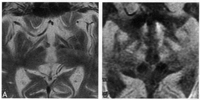
  • fig 2.
    • Download figure
    • Open in new tab
    • Download powerpoint
    fig 2.

    41-year-old male control subject.

    A, Axial T2-weighted image (3540/105) of the midbrain barely depicts the substantia nigra (arrow). The red nucleus (arrowhead) is depicted as a round area of low intensity resulting from the accumulation of iron.

    B, Diffusion-weighted image with a left-right direction diffusion-sensitizing gradient shows a distinct crescent of low signal intensity (arrow) between the cerebral peduncle and the tegmentum. Large arrowhead indicates substantia nigra; small arrowhead, mammillary body.

  • fig 3.
    • Download figure
    • Open in new tab
    • Download powerpoint
    fig 3.

    Thickness of the substantia nigra in the control subjects, the patients with Parkinson's disease, and the patients with secondary parkinsonism. The mean (± SD) thickness of the substantia nigra in the control group (n = 72) was 5.1 ± 0.89 mm, that in the group with Parkinson's disease (n = 47), 4.8 ± 0.75 mm; and that in the group with secondary parkinsonism (n = 10), 3.4 ± 0.53 mm. There was a significant difference between the group with Parkinson's disease and the group with secondary parkinsonism by unpaired Student's t-test (P < .01)

  • fig 4.
    • Download figure
    • Open in new tab
    • Download powerpoint
    fig 4.

    67-year-old man with Parkinson's disease (Hoehn and Yahr scale, 4).

    A, On T2-weighted image (3540/105), the substantia nigra is difficult to identify. There are no abnormal signal changes in the midbrain.

    B, On diffusion-weighted image, the substantia nigra (arrow) is depicted clearly as a crescent of low signal intensity. No atrophy of the substantia nigra is seen.

  • fig 5.
    • Download figure
    • Open in new tab
    • Download powerpoint
    fig 5.

    19-year-old man with secondary parkinsonism (carbon monoxide intoxication) 3 years after onset.

    A, On T2-weighted axial image (3540/105), a hyperintense lesion (arrow) is observed in the globus pallidus; a result of carbon monoxide poisoning.

    B, Diffusion-weighted axial image of the midbrain shows atrophy of both substantia nigra (arrow). The signal intensity is relatively higher than that in the control subjects.

  • fig 6.
    • Download figure
    • Open in new tab
    • Download powerpoint
    fig 6.

    75-year-old man with secondary parkinsonism after bilateral putaminal hemorrhage.

    A, T2-weighted image shows a large hyperintense area in the left putamen and a slitlike hyperintensity in the right putamen. Multiple spotty areas of hyperintensity are also seen in both putamen. The margins of the large area of hyperintensity and of the slitlike hyperintensity are hypointense owing to susceptibility effects resulting from the accumulation of iron (hemosiderin).

    B, On T2-weighted image (3540/105) of the midbrain, the substantia nigra is not distinguishable. There are no abnormal signal changes in the midbrain.

    C, On diffusion-weighted image, both substantia nigra are reduced in size. This image shows hyperintensity in the substantia nigra, which might reflect neuronal degeneration and gliosis.

  • fig 7.
    • Download figure
    • Open in new tab
    • Download powerpoint
    fig 7.

    65-year-old man with progressive supranuclear palsy.

    A, T2-weighted image (3540/105) shows atrophic changes of the midbrain; however, no abnormal signal intensity is evident.

    B, Diffusion-weighted image shows the substantia nigra to be reduced in size.

PreviousNext
Back to top

In this issue

American Journal of Neuroradiology
Vol. 20, Issue 8
1 Sep 1999
  • Table of Contents
  • Index by author
Advertisement
Print
Download PDF
Email Article

Thank you for your interest in spreading the word on American Journal of Neuroradiology.

NOTE: We only request your email address so that the person you are recommending the page to knows that you wanted them to see it, and that it is not junk mail. We do not capture any email address.

Enter multiple addresses on separate lines or separate them with commas.
Evaluation of the Substantia Nigra in Patients with Parkinsonian Syndrome Accomplished Using Multishot Diffusion-Weighted MR Imaging
(Your Name) has sent you a message from American Journal of Neuroradiology
(Your Name) thought you would like to see the American Journal of Neuroradiology web site.
CAPTCHA
This question is for testing whether or not you are a human visitor and to prevent automated spam submissions.
Cite this article
Michito Adachi, Takaaki Hosoya, Tamami Haku, Koichi Yamaguchi, Toru Kawanami
Evaluation of the Substantia Nigra in Patients with Parkinsonian Syndrome Accomplished Using Multishot Diffusion-Weighted MR Imaging
American Journal of Neuroradiology Sep 1999, 20 (8) 1500-1506;

Citation Manager Formats

  • BibTeX
  • Bookends
  • EasyBib
  • EndNote (tagged)
  • EndNote 8 (xml)
  • Medlars
  • Mendeley
  • Papers
  • RefWorks Tagged
  • Ref Manager
  • RIS
  • Zotero
0 Responses
Respond to this article
Share
Bookmark this article
Evaluation of the Substantia Nigra in Patients with Parkinsonian Syndrome Accomplished Using Multishot Diffusion-Weighted MR Imaging
Michito Adachi, Takaaki Hosoya, Tamami Haku, Koichi Yamaguchi, Toru Kawanami
American Journal of Neuroradiology Sep 1999, 20 (8) 1500-1506;
del.icio.us logo Twitter logo Facebook logo Mendeley logo
  • Tweet Widget
  • Facebook Like
  • Google Plus One

Jump to section

  • Article
    • Abstract
    • Methods
    • Results
    • Discussion
    • Conclusion
    • Footnotes
    • References
  • Figures & Data
  • Info & Metrics
  • Responses
  • References
  • PDF

Related Articles

  • No related articles found.
  • PubMed
  • Google Scholar

Cited By...

  • Distinguishing Neuroimaging Features in Patients Presenting with Visual Hallucinations
  • Structural Brain Abnormalities in Patients with Parkinson Disease: A Comparative Voxel-Based Analysis Using T1-Weighted MR Imaging and Magnetization Transfer Imaging
  • Diffusion-weighted MRI differentiates the Parkinson variant of multiple system atrophy from PD
  • Crossref
  • Google Scholar

This article has not yet been cited by articles in journals that are participating in Crossref Cited-by Linking.

More in this TOC Section

  • White Matter Alterations in the Brains of Patients with Active, Remitted, and Cured Cushing Syndrome: A DTI Study
  • Qualitative and Quantitative Analysis of MR Imaging Findings in Patients with Middle Cerebral Artery Stroke Implanted with Mesenchymal Stem Cells
  • Fast Contrast-Enhanced 4D MRA and 4D Flow MRI Using Constrained Reconstruction (HYPRFlow): Potential Applications for Brain Arteriovenous Malformations
Show more Brain

Similar Articles

Advertisement

Indexed Content

  • Current Issue
  • Accepted Manuscripts
  • Article Preview
  • Past Issues
  • Editorials
  • Editor's Choice
  • Fellows' Journal Club
  • Letters to the Editor
  • Video Articles

Cases

  • Case Collection
  • Archive - Case of the Week
  • Archive - Case of the Month
  • Archive - Classic Case

More from AJNR

  • Trainee Corner
  • Imaging Protocols
  • MRI Safety Corner
  • Book Reviews

Multimedia

  • AJNR Podcasts
  • AJNR Scantastics

Resources

  • Turnaround Time
  • Submit a Manuscript
  • Submit a Video Article
  • Submit an eLetter to the Editor/Response
  • Manuscript Submission Guidelines
  • Statistical Tips
  • Fast Publishing of Accepted Manuscripts
  • Graphical Abstract Preparation
  • Imaging Protocol Submission
  • Evidence-Based Medicine Level Guide
  • Publishing Checklists
  • Author Policies
  • Become a Reviewer/Academy of Reviewers
  • News and Updates

About Us

  • About AJNR
  • Editorial Board
  • Editorial Board Alumni
  • Alerts
  • Permissions
  • Not an AJNR Subscriber? Join Now
  • Advertise with Us
  • Librarian Resources
  • Feedback
  • Terms and Conditions
  • AJNR Editorial Board Alumni

American Society of Neuroradiology

  • Not an ASNR Member? Join Now

© 2025 by the American Society of Neuroradiology All rights, including for text and data mining, AI training, and similar technologies, are reserved.
Print ISSN: 0195-6108 Online ISSN: 1936-959X

Powered by HighWire