
Article Figures & Data
Figures
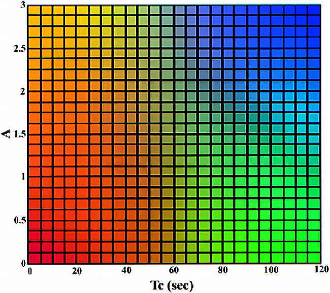
- fig 1.
Two-dimensional color table. The value of the amplitude (A) is from 0 to 3 and that of the distribution time (Tc) is from 0 to 120. The four-corner colors of the color table are yellow, red, blue, and green. The colors of other elements are generated using bilinear interpolation according to their position on the color table. The color mapping of the ROI is determined on the basis of the value of A and Tc in each pixel. Note that at values above 3, A is set to 3; at values above 120, Tc is set to 120
- fig 2.
RSI indices (mean ± 1 standard error) for recurrent IP (open bullets), postoperative changes (solid bullets), and mouth-floor muscle (inverted pyramid) are shown. Note the differences in the magnitude and time course of enhancement. Both types of lesions enhance to a similar final magnitude, but there are marked differences in relative enhancement immediately after contrast injection (P < .01 by Student's t test or Wilcoxon rank sums test)
- fig 3.
A case of postoperative recurrent IP. Patient 2 was referred because of the presence of a polypoid mass in the left superior maxillary sinus wall. This patient had a left nasal fossa IP, which was initially treated with en bloc excision by lateral rhinotomy-medial maxillectomy 15 months before the study was conducted.
A, Conventional MR images (upper left, T1-weighted image [600/20/2]; upper right, PD-weighted image [2500/18/1]; lower left, T2-weighted image [2500/90/1]; lower right, postcontrast T1-weighted image). The T1-weighted image shows isointensity with muscle. The T2-weighted image shows an area of homogeneous hyperintensity in the left superior maxillary sinus wall; the signal intensity is isointense to CSF. Postcontrast studies show homogeneously marked enhancement as compared with enhanced mucosa. The diagnosis of the conventional images is “postoperative changes.”
B, ROI analysis shows a color-coded image suggestive of recurrence.
C, Two-parameter scatter diagram shows amplitude (A) plotted against the distribution time (Tc). The A and Tc of the pixels corresponding to the ROI are shown.
- fig 4.
A case of postoperative changes. Patient 6 was referred because of the presence of a polypoid mass in right maxillary sinus wall. This patient had a right nasal fossa and maxillary sinus IP that was treated initially with en bloc excision via lateral rhinotomy-medial maxillectomy 9 months before the study was conducted.
A, The conventional images are presented in the same order as those shown in figure 3A. The T1-weighted image shows isointensity to muscle. The T2-weighted image shows a region of inhomogeneous hyperintensity in right maxillary sinus wall; the signal intensity of most areas (arrows) in this region is hypointense to CSF and hyperintense to muscle. Postcontrast studies with fat saturation show inhomogeneous, moderate enhancement as compared with enhanced mucosa. The diagnosis of the conventional images is “recurrence.”
B, ROI analysis shows a color-coded zone suggestive of postoperative changes.
C, The A and Tc of the pixels corresponding to the ROI are shown.
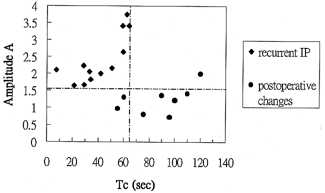
- fig 5.
Two-parameter scatter diagram showing amplitude (A) plotted against the distribution time (Tc). The recurrent IP (diamonds) are localized in the left upper corner, displaying a combination of high amplitude and short distribution time. Tumor recurrence can be defined by a combination of amplitude greater than 1.6 arbitrary units and distribution time less than 65 seconds. The postoperative changes (solid bullets) are characterized by low amplitude (<1.6 arbitrary units) and long distribution time (>65 seconds). Only one postoperatively changed lesion displayed high amplitude. In this case, the lesion still can be distinguished from a recurrent tumor on the basis of distribution time; however, if only the amplitude were considered, the lesion would be misclassified as a tumor


Tables
 TABLE 1:
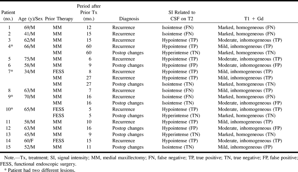
Characteristics, imaging results, and final diagnoses of patients with suspected recurrence of inverted papilloma
- TABLE 2:
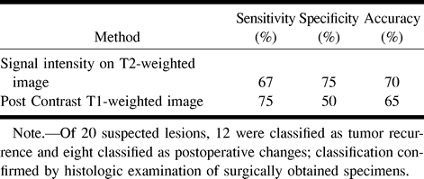
Efficacy of signal intensity on T2-weighted images and postcontrast T1-weighted images for detecting recurrent inverted papilloma
- TABLE 3:
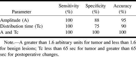
Efficacy of pharmacokinetic parameters for differentiating recurrent tumors from postoperative changes
In this issue


Jump to section
Related Articles
Cited By...
- No citing articles found.