Advertisement

Main menu

  • Home
  • Content
    • Current Issue
    • Accepted Manuscripts
    • Article Preview
    • Past Issue Archive
    • Video Articles
    • AJNR Case Collection
    • Case of the Week Archive
    • Case of the Month Archive
    • Classic Case Archive
  • Special Collections
    • AJNR Awards
    • Low-Field MRI
    • Alzheimer Disease
    • ASNR Foundation Special Collection
    • Photon-Counting CT
    • View All
  • Multimedia
    • AJNR Podcasts
    • AJNR SCANtastic
    • Trainee Corner
    • MRI Safety Corner
    • Imaging Protocols
  • For Authors
    • Submit a Manuscript
    • Submit a Video Article
    • Submit an eLetter to the Editor/Response
    • Manuscript Submission Guidelines
    • Statistical Tips
    • Fast Publishing of Accepted Manuscripts
    • Graphical Abstract Preparation
    • Imaging Protocol Submission
    • Author Policies
  • About Us
    • About AJNR
    • Editorial Board
    • Editorial Board Alumni
  • More
    • Become a Reviewer/Academy of Reviewers
    • Subscribers
    • Permissions
    • Alerts
    • Feedback
    • Advertisers
    • ASNR Home

User menu

  • Alerts
  • Log in

Search

  • Advanced search
American Journal of Neuroradiology

American Journal of Neuroradiology

ASHNR American Society of Functional Neuroradiology ASHNR American Society of Pediatric Neuroradiology ASSR
  • Alerts
  • Log in

Advanced Search

  • Home
  • Content
    • Current Issue
    • Accepted Manuscripts
    • Article Preview
    • Past Issue Archive
    • Video Articles
    • AJNR Case Collection
    • Case of the Week Archive
    • Case of the Month Archive
    • Classic Case Archive
  • Special Collections
    • AJNR Awards
    • Low-Field MRI
    • Alzheimer Disease
    • ASNR Foundation Special Collection
    • Photon-Counting CT
    • View All
  • Multimedia
    • AJNR Podcasts
    • AJNR SCANtastic
    • Trainee Corner
    • MRI Safety Corner
    • Imaging Protocols
  • For Authors
    • Submit a Manuscript
    • Submit a Video Article
    • Submit an eLetter to the Editor/Response
    • Manuscript Submission Guidelines
    • Statistical Tips
    • Fast Publishing of Accepted Manuscripts
    • Graphical Abstract Preparation
    • Imaging Protocol Submission
    • Author Policies
  • About Us
    • About AJNR
    • Editorial Board
    • Editorial Board Alumni
  • More
    • Become a Reviewer/Academy of Reviewers
    • Subscribers
    • Permissions
    • Alerts
    • Feedback
    • Advertisers
    • ASNR Home
  • Follow AJNR on Twitter
  • Visit AJNR on Facebook
  • Follow AJNR on Instagram
  • Join AJNR on LinkedIn
  • RSS Feeds

AJNR Awards, New Junior Editors, and more. Read the latest AJNR updates

Research ArticleBrain

In Vivo Proton MR Spectroscopy of Untreated and Treated Brain Abscesses

Isabella M. Burtscher and Stig Holtås
American Journal of Neuroradiology June 1999, 20 (6) 1049-1053;
Isabella M. Burtscher
  • Find this author on Google Scholar
  • Find this author on PubMed
  • Search for this author on this site
Stig Holtås
  • Find this author on Google Scholar
  • Find this author on PubMed
  • Search for this author on this site
  • Article
  • Figures & Data
  • Info & Metrics
  • Responses
  • References
  • PDF
Loading

Article Figures & Data

Figures

  • Tables
  • fig 1.
    • Download figure
    • Open in new tab
    • Download powerpoint
    fig 1.

    MR images of patient 1 with a brain abscess.

    A–C, T1-weighted axial spin-echo images (630/17/2) before (A) and after (B) contrast injection, and T2-weighted axial fast spin-echo image (3710/90/1) (C).

    D, The proton spectrum (2D chemical-shift PRESS sequence, 1500/270/2) from the abscess cyst, obtained 40 days after the start of initial antibiotic treatment and 12 days after the start of high-dose antibiotic treatment for suspected brain abscess, shows a single lactate peak at 1.3 ppm.

  • fig 2.
    • Download figure
    • Open in new tab
    • Download powerpoint
    fig 2.

    MR images of patient 2 with a brain abscess.

    A–C, T1-weighted axial spin-echo images (630/17/2) before (A) and after (B) contrast injection, and T2-weighted axial fast spin-echo image (3710/90/1) (C).

    D, The proton spectrum (2D chemical-shift PRESS sequence, 1500/270/2) from the abscess cyst, obtained 1 day after the start of antibiotic treatment, shows succinate (Succ), acetate (Ac), alanine (Ala), lactate (Lac), and amino acids (AA).

    E, At a TE of 135, the resonances at 1.5, 1.3, and 0.9 ppm are inverted, which confirms the assignment to alanine, lactate, and amino acids, respectively.

  • fig 3.
    • Download figure
    • Open in new tab
    • Download powerpoint
    fig 3.

    MR images of patient 3.

    A–C, T1-weighted axial spin-echo images (630/17/2) before (A) and after (B) contrast injection, and T2-weighted axial fast spin-echo image (3710/90/1) (C).

    D, Image shows the area of the spectroscopy measurement and the placement of the voxel representing the spectrum (see E).

    E, The proton spectrum (2D chemical-shift PRESS sequence, 1500/270/2) from the abscess cyst shows resonances representing succinate (Succ), acetate (Ac), alanine (Ala), lactate (Lac), and amino acids (AA). Visible small resonances of Cho, Cr, and NAA were interpreted to be caused by partial volume effects. A follow-up examination 2 weeks after the first MR spectroscopic measurement and the institution of high-dose antibiotic therapy revealed a dramatic change in the spectrum from the abscess cyst.

    F and G, All resonances but lactate disappeared (F), a finding that was confirmed by another two follow-up examinations 9 days (G) and 84 days later.

Tables

  • Figures
  • Table1

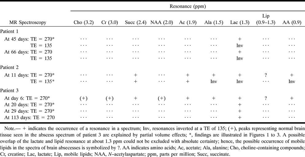
    MR spectroscopic results in three patients

PreviousNext
Back to top

In this issue

American Journal of Neuroradiology
Vol. 20, Issue 6
1 Jun 1999
  • Table of Contents
  • Index by author
Advertisement
Print
Download PDF
Email Article

Thank you for your interest in spreading the word on American Journal of Neuroradiology.

NOTE: We only request your email address so that the person you are recommending the page to knows that you wanted them to see it, and that it is not junk mail. We do not capture any email address.

Enter multiple addresses on separate lines or separate them with commas.
In Vivo Proton MR Spectroscopy of Untreated and Treated Brain Abscesses
(Your Name) has sent you a message from American Journal of Neuroradiology
(Your Name) thought you would like to see the American Journal of Neuroradiology web site.
CAPTCHA
This question is for testing whether or not you are a human visitor and to prevent automated spam submissions.
Cite this article
Isabella M. Burtscher, Stig Holtås
In Vivo Proton MR Spectroscopy of Untreated and Treated Brain Abscesses
American Journal of Neuroradiology Jun 1999, 20 (6) 1049-1053;

Citation Manager Formats

  • BibTeX
  • Bookends
  • EasyBib
  • EndNote (tagged)
  • EndNote 8 (xml)
  • Medlars
  • Mendeley
  • Papers
  • RefWorks Tagged
  • Ref Manager
  • RIS
  • Zotero
0 Responses
Respond to this article
Share
Bookmark this article
In Vivo Proton MR Spectroscopy of Untreated and Treated Brain Abscesses
Isabella M. Burtscher, Stig Holtås
American Journal of Neuroradiology Jun 1999, 20 (6) 1049-1053;
del.icio.us logo Twitter logo Facebook logo Mendeley logo
  • Tweet Widget
  • Facebook Like
  • Google Plus One

Jump to section

  • Article
    • Abstract
    • Case Reports
    • Discussion
    • Acknowledgments
    • Footnotes
    • References
  • Figures & Data
  • Info & Metrics
  • Responses
  • References
  • PDF

Related Articles

  • No related articles found.
  • PubMed
  • Google Scholar

Cited By...

  • Multiparametric imaging in the evaluation of intracerebral abscesses
  • Multiparametric imaging in the evaluation of intracerebral abscesses
  • Intracranial hydatid cyst: imaging findings of a rare disease
  • Role of imaging in the diagnosis of acute bacterial meningitis and its complications
  • In Vivo Proton MR Spectroscopy Evaluation of Pyogenic Brain Abscesses: A Report of 194 Cases
  • Crossref
  • Google Scholar

This article has not yet been cited by articles in journals that are participating in Crossref Cited-by Linking.

More in this TOC Section

  • Usefulness of Quantitative Susceptibility Mapping for the Diagnosis of Parkinson Disease
  • White Matter Alterations in the Brains of Patients with Active, Remitted, and Cured Cushing Syndrome: A DTI Study
  • Qualitative and Quantitative Analysis of MR Imaging Findings in Patients with Middle Cerebral Artery Stroke Implanted with Mesenchymal Stem Cells
Show more Brain

Similar Articles

Advertisement

Indexed Content

  • Current Issue
  • Accepted Manuscripts
  • Article Preview
  • Past Issues
  • Editorials
  • Editor's Choice
  • Fellows' Journal Club
  • Letters to the Editor
  • Video Articles

Cases

  • Case Collection
  • Archive - Case of the Week
  • Archive - Case of the Month
  • Archive - Classic Case

More from AJNR

  • Trainee Corner
  • Imaging Protocols
  • MRI Safety Corner
  • Book Reviews

Multimedia

  • AJNR Podcasts
  • AJNR Scantastics

Resources

  • Turnaround Time
  • Submit a Manuscript
  • Submit a Video Article
  • Submit an eLetter to the Editor/Response
  • Manuscript Submission Guidelines
  • Statistical Tips
  • Fast Publishing of Accepted Manuscripts
  • Graphical Abstract Preparation
  • Imaging Protocol Submission
  • Evidence-Based Medicine Level Guide
  • Publishing Checklists
  • Author Policies
  • Become a Reviewer/Academy of Reviewers
  • News and Updates

About Us

  • About AJNR
  • Editorial Board
  • Editorial Board Alumni
  • Alerts
  • Permissions
  • Not an AJNR Subscriber? Join Now
  • Advertise with Us
  • Librarian Resources
  • Feedback
  • Terms and Conditions
  • AJNR Editorial Board Alumni

American Society of Neuroradiology

  • Not an ASNR Member? Join Now

© 2025 by the American Society of Neuroradiology All rights, including for text and data mining, AI training, and similar technologies, are reserved.
Print ISSN: 0195-6108 Online ISSN: 1936-959X

Powered by HighWire