
Article Figures & Data
Figures
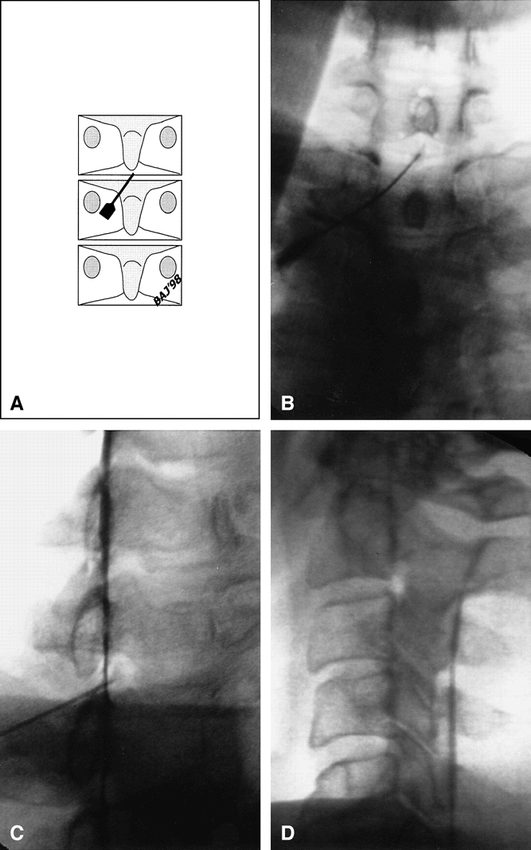
- fig 1.
A, Fluoroscopic image with approximately 15° left and caudal angulation shows a 3.5-inch spinal needle (5-inch needles are used for obese patients), which is advanced toward the superior aspect of the spinal lamina just below the interlaminar gap. Intermittent fluoroscopic checks of position are used after needle advancement. Allowing the needle to contact the superior aspect of the lamina provides depth control.
B, The needle is then redirected cephalad using bevel rotation and control to guide it over the lamina, passing through the ligamentum flavum and into the posterior epidural space at the midline.
C, Schematic representation of needle orientation relative to the posterior elements after advancing the needle tip over the lamina (oblique view).
D, Anteroposterior fluoroscopic image shows the orientation of the needle as it is advanced over the lamina from a left posterolateral and slightly caudal approach.
E, The needle is then advanced into the epidural space and contrast agent is injected after negative testing for CSF aspiration. On this image, the tip of the needle is seen in the midline and contrast material is beginning to flow into the epidural space. A total of 5 to 6 mL of iohexol, 240 mg/mL, are injected under direct fluoroscopic observation to ensure distribution within the epidural space and exclude opacification of the thecal sac (subarachnoid space), venous structures, or adjacent compartments (subdural/extraarachnoid or paraspinous). After confirmation of epidural space opacification and filming, 2 to 3 mL of betamethasone sodium phosphate/betamethasone acetate suspension followed by 3 to 5 mL of 1% lidocaine or 0.5% miconazole nitrate are injected through the same needle, which has not been moved.
- fig 2.
A and B, Anteroposterior (A) and lateral (B) radiographs after contrast injection. The needle is positioned centrally within the dorsal epidural space using technique 1 or 2; the contrast material is seen circumferentially within the epidural space and along the proximal nerve sheaths (arrows).
fig 3. Schematic representation of needle placement and orientation relative to osseous structures. The needle is aligned with the X-ray beam and is seen en face at fluoroscopy 15° to 20° off midline. A 22-gauge epidural needle is passed in a coaxial fashion through the 18-gauge introducer needle and into the dorsal epidural space at the midline. Anteroposterior and lateral fluoroscopy is used to confirm midline needle tip position in the dorsal epidural space.
- fig 4.
A, After sterile preparation and draping, a 22-gauge spinal needle is carefully advanced into the appropriate neuroforamen immediately subjacent to the vertebral pedicle via a dorsal/lateral approach using intermittent, brief (1–2 second), low-dose fluoroscopic checks. Oblique radiograph after contrast injection shows the needle tip within the right L5–S1 neural foramen. There is opacification of the proximal L5 nerve sheath, with epidural reflux of contrast material, which extends from the L4–L5 level to the S1–S2 level within the epidural space. Ideal needle placement produces opacification of the proximal nerve sheath and adjacent epidural space. After filming (anteroposterior, lateral, and/or oblique images), 2 to 3 mL of water-soluble steroid mixture followed by 3 to 5 mL of local anesthetic are injected through the same needle. Postinjection films are then obtained to document dispersal of the injectate.
B, Schematic representation depicts position of the needle within the foramen using a posterolateral approach. The target for the needle tip in this position is the inferior aspect of the pedicle, which defines the superior margin of the neural foramen. The tip should be at the central (six-o'clock) or slightly medial position for a transforaminal epidural injection. It is positioned slightly lateral to this for a selective nerve root injection.
fig 5. Anteroposterior radiograph after contrast injection via a right S1 transforaminal epidural approach reveals contrast material extending along the S1 and S2 nerve root sheaths and within the sacral epidural space on the right. The contrast material does not ascend above the L5–S1 level owing to a large herniated nucleus pulposus, which was documented on a CT study.
- fig 6.
A, Schematic representation of anteroposterior view shows the orientation and position of the needle relative to the sacrum. The needle is passed into the sacral hiatus from below and posteriorly, but is not advanced above the S3 level to avoid inadvertent thecal sac puncture.
B and C, Anteroposterior (B) and lateral (C) radiographs after needle placement in the midline through the sacral hiatus reveal contrast material throughout the sacral epidural space (5 to 6 mL of contrast agent is injected before the radiographs are obtained). After documenting opacification of the caudal epidural space and excluding venous or intrathecal opacification, 2 to 3 mL of steroid mixture followed by 3 to 5 mL of local anesthetic are introduced. Postinjection films are obtained to document dispersal of the injectate. Note that contrast material extends cephalad to the L5–S1 level, but not above this. A significant amount of injectate remains in the caudal sacral canal.
- fig 7.
A, Schematic representation of an anteroposterior view shows orientation of the blunt-tipped Whitacre needle relative to the lamina at the C7–T1 level. The needle tip is midline in the dorsal epidural space. An introducer needle is used before placement of the blunt-tipped 22-gauge Whitacre needle. In smaller patients, the introducer needle may be left in place for coaxial insertion of the needle. In larger patients, the 18-gauge needle is removed after skin puncture and before introduction of the Whitacre needle, as the hub of this needle limits the depth of the epidural needle insertion. The needle is advanced rostral and central toward the midline interlaminar gap with the use of intermittent fluoroscopy. Contact with the lamina before entry into the epidural space provides depth control, which is crucial to prevent cord injury.
B, After needle placement, 4 to 5 mL of iohexol, 240 mg/mL, is injected, followed by anteroposterior radiography, which reveals diffuse opacification of the lower cervical and upper thoracic epidural space with extension of contrast material along the proximal nerve sheaths bilaterally.
C, Oblique radiograph shows needle placement over the lamina and into the spinal canal. Contrast agent in the posterior epidural space is profiled in this projection. No venous or thecal sac opacification is exhibited.
D, Lateral radiograph after steroid injection and removal of the needle reveals widespread dispersal of the previously injected contrast agent within the cervical epidural space, outlining the unopacified cervical thecal sac.
- fig 8.
A–C, Anteroposterior (A), oblique (B), and lateral (C) radiographs after thoracic interlaminar epidural injection of contrast material reveal opacification of the lower thoracic epidural space dorsally before injection of steroid and local anesthetic. The Whitacre needle tip is in the midline within the posterior epidural space. Contrast material is seen extending along the proximal thoracic nerve sheaths (arrows)


In this issue


Jump to section
Related Articles
Cited By...
- Comparison of the contralateral oblique view with the lateral view for mid-thoracic epidural access under fluoroscopic guidance: a randomized controlled trial
- Use of epidurography in the perioperative and acute pain setting
- Inadvertent Intrafacet Injection during Lumbar Interlaminar Epidural Steroid Injection: A Comparison of CT Fluoroscopic and Conventional Fluoroscopic Guidance
- The Incidence of Lumbar Discectomy after Epidural Steroid Injections or Selective Nerve Root Blocks
- Immediate Pain Response to Interlaminar Lumbar Epidural Steroid Administration: Response Characteristics and Effects of Anesthetic Concentration
- CT Fluoroscopy-Guided Cervical Interlaminar Steroid Injections: Safety, Technique, and Radiation Dose Parameters
- Treatment of Lumbar Disc Herniation: Epidural Steroid Injection Compared with Discectomy: A Prospective, Randomized Study
- The American Journal of Neuroradiology 1980-1999 Where We Have Been: Where We Are Going