
Article Figures & Data
Figures
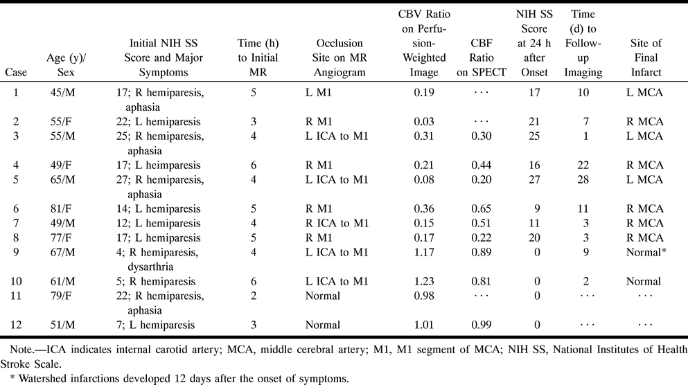
- fig 1.
Case 8: 77-year-old woman with hyperacute ischemic stroke with arterial occlusion and decreased CBV.
A, T2-weighted MR image (3500/90) is normal except for abnormal subtle high signal intensity in right basal ganglia.
B, 3D time-of-flight MR angiogram reveals occlusion at M1 segment of the right MCA.
C, CBV map shows decreased CBV in right MCA distribution.
D, CBV map shows irregular ROIs placed for measurement of CBV ratio and time–signal intensity curves between the region of decreased CBV and contralateral normal region. Calculated CBV ratio was 0.17.
E, Time–signal intensity curves measured during passage of contrast material show no signal change in right MCA distribution compared with normal signal reduction in left MCA distribution.
F, 99mTc-HMPAO brain SPECT scan obtained during the hyperacute stage at approximately the same level as A and C reveals severe hypoperfusion throughout right MCA distribution.
G, Follow-up CT scan 3 days after the onset of symptoms shows well-defined infarction in right MCA distribution, which corresponds to the region of decreased CBV.
- fig 2.
Case 10: 61-year-old man with hyperacute ischemic stroke with arterial occlusion and increased CBV.
A, T2-weighted image (3500/90) is normal except for small chronic lesion with hemosiderin rim in left basal ganglia.
B, 3D time-of-flight MR angiogram reveals occlusion of left internal carotid artery through M1 segment of the left MCA.
C, CBV map shows increased CBV throughout left MCA distribution.
D, CBV map shows irregular ROIs placed for measurement of CBV ratio and time–signal intensity curves between the region of increased CBV and contralateral normal region. Calculated CBV ratio was 1.23.
E, Time–signal intensity curves measured during passage of contrast material show delayed arrival time and delayed washout of the contrast material in left MCA distribution, suggesting collateral flow.
F, 99mTc-HMPAO brain SPECT scan obtained during the hyperacute stage at approximately the same level as A and C reveals mild hypoperfusion throughout left MCA and anterior cerebral artery distribution.
G, Follow-up T2-weighted MR image (3500/90) obtained 2 days after the onset of symptoms is normal except for small chronic lesion in left basal ganglia.


Tables

Summary of findings
In this issue


Jump to section
Related Articles
Cited By...
- Fetal Origin of the Posterior Cerebral Artery Produces Left-Right Asymmetry on Perfusion Imaging
- Management of Stroke in Infants and Children: A Scientific Statement From a Special Writing Group of the American Heart Association Stroke Council and the Council on Cardiovascular Disease in the Young
- Rapid Perfusion Abnormality Estimation in Acute Stroke With Temporal Correlation Analysis
- Relationship between severity of MR perfusion deficit and DWI lesion evolution
- Systematic Review of Diffusion and Perfusion Imaging in Acute Ischemic Stroke
- Prediction of Malignant Middle Cerebral Artery Infarction by Diffusion-Weighted Imaging