
Article Figures & Data
Figures
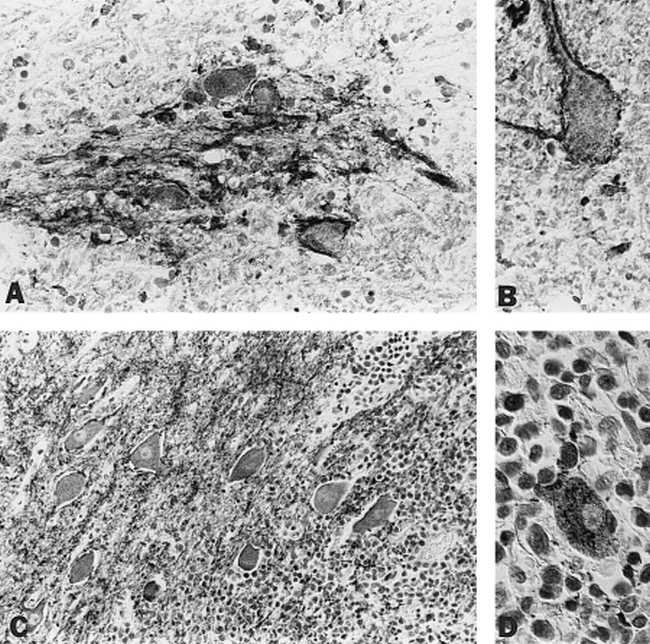
fig 1. Immunohistochemical study† of a normal cervical spinal cord of a 75-year-old man (A and B), and of a dorsal medulla of a 79-year-old woman with malignant lymphoma (C and D).
A, A small cluster of spinal cord neurons, with cellular and superficial synaptophysin reactivity, is embedded in white matter. (original magn. ×330)
B, A cervical spinal cord neuron, synaptophysin-positive, is embedded in white matter. (original magn. ×500)
C, Native neurons of dorsal medulla, synaptophysin-positive, span the borderzone with lymphoma. (original magn. ×330)
D, Neurons are slightly deeper in the tumor. (original magn. ×500)
† For staining methods, see reference 4.

In this issue


Jump to section
Related Articles
Cited By...
- No citing articles found.