
Article Figures & Data
Figures
- fig 1.
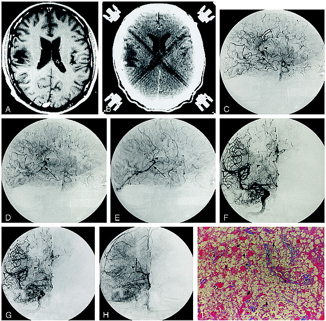
Patient 1.
A, Contrast-enhanced axial T1-weighted MR image (600/14/2) shows an enhancing lesion in the left brachium pontis.
B, Contrast-enhanced CT scan at the same level as the MR image. Although there is considerable streak artifact in the posterior fossa, there is an enhancing lesion in the left brachium pontis (arrow) corresponding to that identified on the MR image.
C–F, Early arterial (C), mid-arterial (D), capillary (E), and venous (F) phase images from the left vertebral artery stereo angiogram (anteroposterior projection) show an abnormal parenchymal blush (double arrows) in the left cerebellar hemisphere corresponding to the lesion seen on the CT and MR studies. There is arteriovenous shunting with early opacification of the left lateral recess and cerebellomedullary veins (curved arrows), which empty into the left sigmoidal sinus.
G, Histopathologic specimen shows exuberant perivascular inflammation (inf). The surrounding tissue shows gliosis (curved arrows) and microglial activation (straight arrows) (hematoxylin-eosin, original magnification ×100).
H, Higher-power view shows the mixed population of inflammatory cells, including lymphocytes, monocytes, and plasma cells (arrowheads). Note the plumping of the endothelial cells (arrow) (hematoxylin-eosin, original magnification ×200).
I, Azocarmine stain of tissue specimen in G shows reduplication of the basal lamina (arrows), seen as a fine connective tissue network (azocarmine stain, original magnification ×200).
- fig 2.
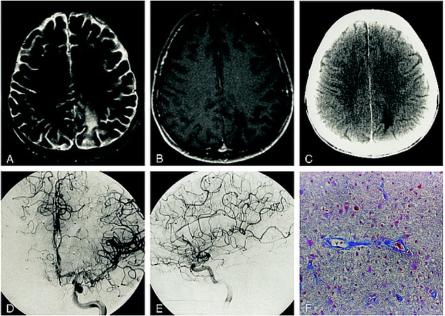
Patient 2.
A, Contrast-enhanced T1-weighted MR image (600/14/2) illustrates the nonenhancing right posterior frontal lesion.
B, Corresponding contrast-enhanced CT scan shows a hypodense deep and subcortical white matter lesion.
C–E, Late arterial (C), capillary (D), and early venous (E) phase digital subtraction angiographic (DSA) images (lateral projection) from the accompanying right internal carotid artery (ICA) angiogram depict a region of arteriovenous shunting corresponding to the location of the MR abnormality. A dense parenchymal blush is evident throughout the inferior frontoparietal region (curved arrow, C). Arteriovenous shunting results in the early opacification of a direct atrial vein (large arrowhead) and the posterior third of the right internal cerebral vein (arrow). An early draining cortical parietal vein is seen in D and E (small arrowhead).
F–H, Corresponding mid-arterial (F), late arterial (G), and early venous (H) phase frontal DSA images confirm early opacification of the atrial (arrowhead) and right internal cerebral (arrow) veins.
I, Histologic section shows several vascular channels (v) without evidence of reduplication of the basal lamina. There is mingling of numerous histiocytes (straight arrows) and reactive astrocytes (curved arrows), characteristic of a demyelinating process. Some of the astrocytic cells have enlarged hyperplastic nuclei (open arrows) often seen in PML (azocarmine stain, original magnification ×100).
- fig 3.
Patient 3.
A and B, Axial T2-weighted (3400/119/1) (A) and contrast-enhanced T1-weighted (600/14/2) (B) MR images show the nonenhancing left frontal lesion.
C, Corresponding contrast-enhanced CT scan shows a nonenhancing hypodense white matter lesion.
D and E, Arterial (D) and venous (E) phase DSA images (lateral projection) from the accompanying left ICA stereo angiogram depict a region of arteriovenous shunting corresponding to the location of the MR abnormality. An area of abnormal parenchymal blush (curved open arrow) is associated with arteriovenous shunting, resulting in the early opacification of an anterior caudate vein (arrowhead), which empties via the thalamostriate trunk into the left internal cerebral vein (straight arrow). Contrast within the vein of Galen is identified during the arterial phase (curved solid arrow, D).
F, Histopathologic specimen from the region of abnormal parenchymal blush and arteriovenous shunting show numerous small vascular channels (v) outlined by azocarmine. The neuropile (arrowheads) is rarified (azocarmine stain, original magnification ×100).
- fig 4.
Patient 5.
A and B, Axial T2-weighted (3400/119/1) (A) and contrast-enhanced T1-weighted (600/14/2) (B) MR images show a nonenhancing left parietal focus of PML.
C, Accompanying contrast-enhanced CT scan shows a nonenhancing hypodensity in the white matter of the left parietal lobe.
D and E, Anteroposterior (D) and lateral (E) arterial phase DSA images from the left ICA stereo angiogram fail to show any abnormal parenchymal blush or evidence of arteriovenous shunting.
F, A vascular channel (v) devoid of perivascular inflammation is outlined by azocarmine, illustrating the ordinary histologic appearance in this patient with an angiographically occult lesion (azocarmine stain, original magnification ×100).


Tables

Imaging findings in six patients with biopsy-proved progressive multifocal leukoencephalopathy


