Advertisement

Main menu

  • Home
  • Content
    • Current Issue
    • Accepted Manuscripts
    • Article Preview
    • Past Issue Archive
    • Video Articles
    • AJNR Case Collection
    • Case of the Week Archive
    • Case of the Month Archive
    • Classic Case Archive
  • Special Collections
    • AJNR Awards
    • Low-Field MRI
    • Alzheimer Disease
    • ASNR Foundation Special Collection
    • Photon-Counting CT
    • View All
  • Multimedia
    • AJNR Podcasts
    • AJNR SCANtastic
    • Trainee Corner
    • MRI Safety Corner
    • Imaging Protocols
  • For Authors
    • Submit a Manuscript
    • Submit a Video Article
    • Submit an eLetter to the Editor/Response
    • Manuscript Submission Guidelines
    • Statistical Tips
    • Fast Publishing of Accepted Manuscripts
    • Graphical Abstract Preparation
    • Imaging Protocol Submission
    • Author Policies
  • About Us
    • About AJNR
    • Editorial Board
    • Editorial Board Alumni
  • More
    • Become a Reviewer/Academy of Reviewers
    • Subscribers
    • Permissions
    • Alerts
    • Feedback
    • Advertisers
    • ASNR Home

User menu

  • Alerts
  • Log in

Search

  • Advanced search
American Journal of Neuroradiology

American Journal of Neuroradiology

ASHNR American Society of Functional Neuroradiology ASHNR American Society of Pediatric Neuroradiology ASSR
  • Alerts
  • Log in

Advanced Search

  • Home
  • Content
    • Current Issue
    • Accepted Manuscripts
    • Article Preview
    • Past Issue Archive
    • Video Articles
    • AJNR Case Collection
    • Case of the Week Archive
    • Case of the Month Archive
    • Classic Case Archive
  • Special Collections
    • AJNR Awards
    • Low-Field MRI
    • Alzheimer Disease
    • ASNR Foundation Special Collection
    • Photon-Counting CT
    • View All
  • Multimedia
    • AJNR Podcasts
    • AJNR SCANtastic
    • Trainee Corner
    • MRI Safety Corner
    • Imaging Protocols
  • For Authors
    • Submit a Manuscript
    • Submit a Video Article
    • Submit an eLetter to the Editor/Response
    • Manuscript Submission Guidelines
    • Statistical Tips
    • Fast Publishing of Accepted Manuscripts
    • Graphical Abstract Preparation
    • Imaging Protocol Submission
    • Author Policies
  • About Us
    • About AJNR
    • Editorial Board
    • Editorial Board Alumni
  • More
    • Become a Reviewer/Academy of Reviewers
    • Subscribers
    • Permissions
    • Alerts
    • Feedback
    • Advertisers
    • ASNR Home
  • Follow AJNR on Twitter
  • Visit AJNR on Facebook
  • Follow AJNR on Instagram
  • Join AJNR on LinkedIn
  • RSS Feeds

AJNR Awards, New Junior Editors, and more. Read the latest AJNR updates

Research ArticleBrain

Ultrafast Diffusion-Sensitive MR Imaging of Brain on an Open Scanner at 0.2 T

Fritz Schick, Thomas Nägele, Dirk Wildgruber, Jochen Trübenbach and Claus D. Claussen
American Journal of Neuroradiology January 1999, 20 (1) 53-61;
Fritz Schick
aFrom the Departments of Diagnostic Radiology (F.S., J.T., C.D.C.), Neuroradiology (T.N.), and Neurology (D.W.), University of Tübingen, Germany.
  • Find this author on Google Scholar
  • Find this author on PubMed
  • Search for this author on this site
Thomas Nägele
aFrom the Departments of Diagnostic Radiology (F.S., J.T., C.D.C.), Neuroradiology (T.N.), and Neurology (D.W.), University of Tübingen, Germany.
  • Find this author on Google Scholar
  • Find this author on PubMed
  • Search for this author on this site
Dirk Wildgruber
aFrom the Departments of Diagnostic Radiology (F.S., J.T., C.D.C.), Neuroradiology (T.N.), and Neurology (D.W.), University of Tübingen, Germany.
  • Find this author on Google Scholar
  • Find this author on PubMed
  • Search for this author on this site
Jochen Trübenbach
aFrom the Departments of Diagnostic Radiology (F.S., J.T., C.D.C.), Neuroradiology (T.N.), and Neurology (D.W.), University of Tübingen, Germany.
  • Find this author on Google Scholar
  • Find this author on PubMed
  • Search for this author on this site
Claus D. Claussen
aFrom the Departments of Diagnostic Radiology (F.S., J.T., C.D.C.), Neuroradiology (T.N.), and Neurology (D.W.), University of Tübingen, Germany.
  • Find this author on Google Scholar
  • Find this author on PubMed
  • Search for this author on this site
  • Article
  • Figures & Data
  • Info & Metrics
  • Responses
  • References
  • PDF
Loading

Article Figures & Data

Figures

  • Tables
  • fig 1.
    • Download figure
    • Open in new tab
    • Download powerpoint
    fig 1.

    Single-shot diffusion-weighted imaging with the SPLICE acquisition technique.

    A, Sequence of RF pulses and magnetic field gradients. Diffusion weighting is achieved by a spin-echo preparation with strong gradients applied before and after the 180° refocusing pulse. The modified fast spin-echo train with two families of signals (E1 and E2) is generated by repetition of the elements inside the box. Two complete magnitude images from echo trains E1 and E2, respectively, should be added to obtain the final image with higher SNR.

    B, Phase-encoding steps for half-Fourier reconstruction from the signals in echo train E1 and E2, respectively. Sixty-four lines in the final image are considered in the given example.

    C, Phase encoding with centric reordering of the steps for an acquisition of a complete raw data matrix with 64 steps. Data recording starts after six cycles in the fast spin-echo train. This delay allows the spin system to reach an equilibrium state with similar signal intensities in both echo families E1 and E2.
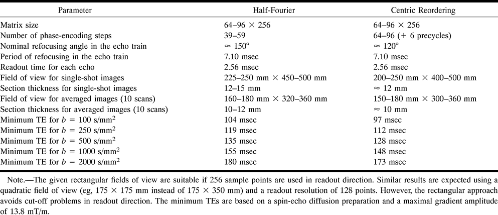
  • fig 2.
    • Download figure
    • Open in new tab
    • Download powerpoint
    fig 2.

    Examples of different types of SPLICE images from the head of a 36-year-old healthy volunteer. Each image, with a 12-mm thickness, was recorded in less than 1 s. The FOV was 210 × 420 mm with a matrix of 96 × 256. A and C were recorded without diffusion gradients, whereas diffusion sensitizing with b = 1000 s/mm2 (maximum amplitude 13.8 mT/m, from 8.0 mT/m in all three orientations simultaneously) was applied for the images in B and D. The timing scheme in the spin-echo preparation remained unchanged for all images. The images recorded with half-Fourier acquisition (A, B) show less blurring artifacts, whereas the SNR is superior in images recorded with centric reordering (C, D). The displayed images are magnified and do not show the entire matrix in read direction.

    A, Half-Fourier recording as shown in figure 1B; 55 phase-encoding steps; TE = 155; no diffusion preparation. SNR of brain is 5.5.

    B, Half-Fourier technique, with diffusion preparation. SNR of brain is 2.0.

    C, Centric reordering as shown in figure 1C; 96 phase-encoding steps; TE = 148; no diffusion preparation. SNR of brain is 7.3.

    D, Centric reordering, with diffusion preparation. SNR of brain is 2.3.
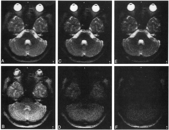
  • fig 3.
    • Download figure
    • Open in new tab
    • Download powerpoint
    fig 3.

    Averaging of several single-shot images allows improved SNR and/or smaller FOV. Averaged transverse sections with a thickness of 10 mm and an FOV of 175 × 350 mm (matrix 96 × 256) from eight single-shot scans in a 28-year-old volunteer. The entire measuring time for one set of images was about 45 s (using a TR of 6 s). Centric reordering and recording of the entire matrix as in figure 1C were applied.

    A, TE = 112; no diffusion sensitivity. SNR of brain is 22.

    B, TE = 112; diffusion preparation with a b value of 250 s/mm2. SNR of brain is 15.

    C, TE = 128; no diffusion sensitivity.

    D, TE = 128; diffusion preparation with a b value of 500 s/mm2.

    E, TE = 148; no diffusion sensitivity. SNR of brain is 15.

    F, TE = 148; diffusion preparation with a b value of 1000 s/mm2. SNR of brain is 3.3.
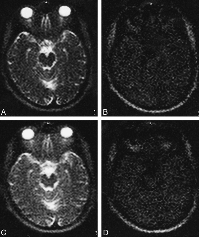
  • fig 4.
    • Download figure
    • Open in new tab
    • Download powerpoint
    fig 4.

    Effect of the addition of two separate magnitude images from echoes E1 and E2 in the modified fast spin-echo train in figure 1A. Coronal single-shot images from a 74-year-old patient with a large ischemic lesion (arrow, F) were recorded using the half-Fourier approach. Section thickness, 12 mm; FOV, 175 × 350 mm; matrix, 96 × 256.

    A–C, TE = 155; no diffusion sensitivity.

    D–F, TE = 155; diffusion preparation with a b value of 1000 s/mm2.

    A and D are images from echoes E1; B and E are images from echoes E2; C and F are the addition of images from E1 and E2. Signal intensity is doubled, whereas noise increases by a factor of Embedded Image. CNR of the lesion versus normal brain is 11 in F.

  • fig 5.
    • Download figure
    • Open in new tab
    • Download powerpoint
    fig 5.

    Transverse images from one section of the brain of a 74-year-old patient with occlusion of the middle cerebral artery, 10 days after onset of stroke symptoms.

    A, Standard T1-weighted fast spin-echo image (400/15/2 [TR/TE/excitations]).

    B, Standard T2-weighted fast spin-echo image (4000/117/1).

    C, Single-scan half-Fourier image. Section thickness, 12 mm; FOV, 250 × 500 mm; matrix, 96 × 256; TE = 155; no diffusion sensitivity.

    D, Single-scan half-Fourier image. Section thickness, 12 mm; FOV, 250 × 500 mm; matrix, 96 × 256; TE = 155; diffusion preparation with a b value of 1000 s/mm2. CNR of the lesion (arrows) versus normal brain is 12.

    E, Ten single-scan images from a set of 20 images were averaged. Half-Fourier approach, section thickness, 10 mm; FOV, 175 × 350 mm; matrix, 96 × 256; TE = 155; no diffusion sensitivity.

    F, Ten single-scan images from a set of 20 images were averaged. Half-Fourier approach, section thickness, 12 mm; FOV, 250 × 500 mm; matrix, 96 × 256; TE = 155; diffusion preparation with a b value of 1000 s/mm2. CNR of the lesion versus normal brain is 13.
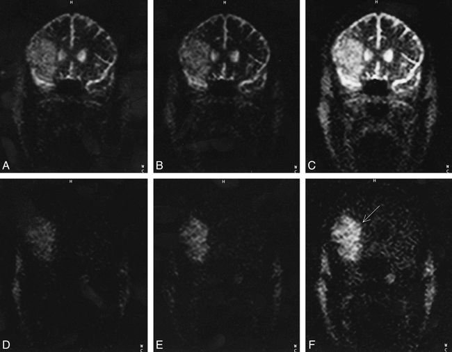
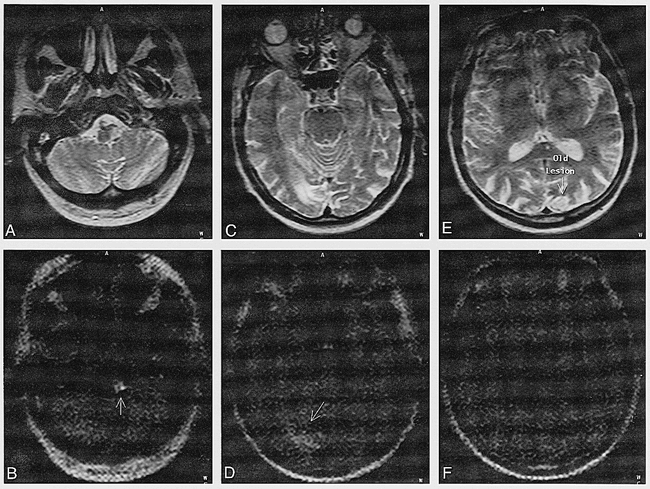
  • fig 6.
    • Download figure
    • Open in new tab
    • Download powerpoint
    fig 6.

    Clinical diffusion imaging in a 58-year-old patient with ischemic brain insults of different ages.

    A, C, E, Anatomic details are well depicted on T2-weighted standard fast spin-echo images (4000/106/1).

    B, D, F, Images show averaged signals from six single-shot scans with half-Fourier reconstruction. Diffusion-weighting (b = 1000 s/mm2, TE = 155) allows distinction between the relatively recent lesion (6 days old, with markedly restricted diffusion (CNR = 10; arrow, B), the older lesion (4 weeks old) with slight signal enhancement (CNR = 4; arrow, D), and the 1-year-old lesion without any visible diffusion abnormalities. FOV, 175 × 350 mm; matrix, 96 × 256; section thickness, 10 mm.

Tables

  • Figures
  • Table1

    Suitable parameters of sequences with half-Fourier reconstruction versus centric reordering of phase-encoding for brain diffusion imaging at 0.2 T

PreviousNext
Back to top

In this issue

American Journal of Neuroradiology
Vol. 20, Issue 1
1 Jan 1999
  • Table of Contents
  • Index by author
Advertisement
Print
Download PDF
Email Article

Thank you for your interest in spreading the word on American Journal of Neuroradiology.

NOTE: We only request your email address so that the person you are recommending the page to knows that you wanted them to see it, and that it is not junk mail. We do not capture any email address.

Enter multiple addresses on separate lines or separate them with commas.
Ultrafast Diffusion-Sensitive MR Imaging of Brain on an Open Scanner at 0.2 T
(Your Name) has sent you a message from American Journal of Neuroradiology
(Your Name) thought you would like to see the American Journal of Neuroradiology web site.
CAPTCHA
This question is for testing whether or not you are a human visitor and to prevent automated spam submissions.
Cite this article
Fritz Schick, Thomas Nägele, Dirk Wildgruber, Jochen Trübenbach, Claus D. Claussen
Ultrafast Diffusion-Sensitive MR Imaging of Brain on an Open Scanner at 0.2 T
American Journal of Neuroradiology Jan 1999, 20 (1) 53-61;

Citation Manager Formats

  • BibTeX
  • Bookends
  • EasyBib
  • EndNote (tagged)
  • EndNote 8 (xml)
  • Medlars
  • Mendeley
  • Papers
  • RefWorks Tagged
  • Ref Manager
  • RIS
  • Zotero
0 Responses
Respond to this article
Share
Bookmark this article
Ultrafast Diffusion-Sensitive MR Imaging of Brain on an Open Scanner at 0.2 T
Fritz Schick, Thomas Nägele, Dirk Wildgruber, Jochen Trübenbach, Claus D. Claussen
American Journal of Neuroradiology Jan 1999, 20 (1) 53-61;
del.icio.us logo Twitter logo Facebook logo Mendeley logo
  • Tweet Widget
  • Facebook Like
  • Google Plus One

Jump to section

  • Article
    • Abstract
    • Methods
    • Results
    • Discussion
    • Acknowledgments
    • Footnotes
    • References
  • Figures & Data
  • Info & Metrics
  • Responses
  • References
  • PDF

Related Articles

  • No related articles found.
  • PubMed
  • Google Scholar

Cited By...

  • No citing articles found.
  • Crossref
  • Google Scholar

This article has not yet been cited by articles in journals that are participating in Crossref Cited-by Linking.

More in this TOC Section

  • Optimal MRI Sequence for Identifying Occlusion Location in Acute Stroke: Which Value of Time-Resolved Contrast-Enhanced MRA?
  • Evaluating the Effects of White Matter Multiple Sclerosis Lesions on the Volume Estimation of 6 Brain Tissue Segmentation Methods
  • Quiet PROPELLER MRI Techniques Match the Quality of Conventional PROPELLER Brain Imaging Techniques
Show more Brain

Similar Articles

Advertisement

Indexed Content

  • Current Issue
  • Accepted Manuscripts
  • Article Preview
  • Past Issues
  • Editorials
  • Editor's Choice
  • Fellows' Journal Club
  • Letters to the Editor
  • Video Articles

Cases

  • Case Collection
  • Archive - Case of the Week
  • Archive - Case of the Month
  • Archive - Classic Case

More from AJNR

  • Trainee Corner
  • Imaging Protocols
  • MRI Safety Corner
  • Book Reviews

Multimedia

  • AJNR Podcasts
  • AJNR Scantastics

Resources

  • Turnaround Time
  • Submit a Manuscript
  • Submit a Video Article
  • Submit an eLetter to the Editor/Response
  • Manuscript Submission Guidelines
  • Statistical Tips
  • Fast Publishing of Accepted Manuscripts
  • Graphical Abstract Preparation
  • Imaging Protocol Submission
  • Evidence-Based Medicine Level Guide
  • Publishing Checklists
  • Author Policies
  • Become a Reviewer/Academy of Reviewers
  • News and Updates

About Us

  • About AJNR
  • Editorial Board
  • Editorial Board Alumni
  • Alerts
  • Permissions
  • Not an AJNR Subscriber? Join Now
  • Advertise with Us
  • Librarian Resources
  • Feedback
  • Terms and Conditions
  • AJNR Editorial Board Alumni

American Society of Neuroradiology

  • Not an ASNR Member? Join Now

© 2025 by the American Society of Neuroradiology All rights, including for text and data mining, AI training, and similar technologies, are reserved.
Print ISSN: 0195-6108 Online ISSN: 1936-959X

Powered by HighWire