Advertisement

Main menu

  • Home
  • Content
    • Current Issue
    • Accepted Manuscripts
    • Article Preview
    • Past Issue Archive
    • Video Articles
    • AJNR Case Collection
    • Case of the Week Archive
    • Case of the Month Archive
    • Classic Case Archive
  • Special Collections
    • AJNR Awards
    • Low-Field MRI
    • Alzheimer Disease
    • ASNR Foundation Special Collection
    • Photon-Counting CT
    • View All
  • Multimedia
    • AJNR Podcasts
    • AJNR SCANtastic
    • Trainee Corner
    • MRI Safety Corner
    • Imaging Protocols
  • For Authors
    • Submit a Manuscript
    • Submit a Video Article
    • Submit an eLetter to the Editor/Response
    • Manuscript Submission Guidelines
    • Statistical Tips
    • Fast Publishing of Accepted Manuscripts
    • Graphical Abstract Preparation
    • Imaging Protocol Submission
    • Author Policies
  • About Us
    • About AJNR
    • Editorial Board
    • Editorial Board Alumni
  • More
    • Become a Reviewer/Academy of Reviewers
    • Subscribers
    • Permissions
    • Alerts
    • Feedback
    • Advertisers
    • ASNR Home

User menu

  • Alerts
  • Log in

Search

  • Advanced search
American Journal of Neuroradiology

American Journal of Neuroradiology

ASHNR American Society of Functional Neuroradiology ASHNR American Society of Pediatric Neuroradiology ASSR
  • Alerts
  • Log in

Advanced Search

  • Home
  • Content
    • Current Issue
    • Accepted Manuscripts
    • Article Preview
    • Past Issue Archive
    • Video Articles
    • AJNR Case Collection
    • Case of the Week Archive
    • Case of the Month Archive
    • Classic Case Archive
  • Special Collections
    • AJNR Awards
    • Low-Field MRI
    • Alzheimer Disease
    • ASNR Foundation Special Collection
    • Photon-Counting CT
    • View All
  • Multimedia
    • AJNR Podcasts
    • AJNR SCANtastic
    • Trainee Corner
    • MRI Safety Corner
    • Imaging Protocols
  • For Authors
    • Submit a Manuscript
    • Submit a Video Article
    • Submit an eLetter to the Editor/Response
    • Manuscript Submission Guidelines
    • Statistical Tips
    • Fast Publishing of Accepted Manuscripts
    • Graphical Abstract Preparation
    • Imaging Protocol Submission
    • Author Policies
  • About Us
    • About AJNR
    • Editorial Board
    • Editorial Board Alumni
  • More
    • Become a Reviewer/Academy of Reviewers
    • Subscribers
    • Permissions
    • Alerts
    • Feedback
    • Advertisers
    • ASNR Home
  • Follow AJNR on Twitter
  • Visit AJNR on Facebook
  • Follow AJNR on Instagram
  • Join AJNR on LinkedIn
  • RSS Feeds

AJNR Awards, New Junior Editors, and more. Read the latest AJNR updates

Research ArticlePEDIATRIC NEUROIMAGING

Clinical Characteristics of Branchial Cleft Anomalies

Neil Rowlands, Catherine Russo, Kevin Liu, Anthony Yassall, Tendy Chiang and Mai-Lan Ho
American Journal of Neuroradiology March 2025, DOI: https://doi.org/10.3174/ajnr.A8604
Neil Rowlands
aFrom the Ohio University College of Osteopathic Medicine (N.R., A.Y.), Dublin, Ohio
  • Find this author on Google Scholar
  • Find this author on PubMed
  • Search for this author on this site
  • ORCID record for Neil Rowlands
Catherine Russo
bThe Ohio State University (C.R.), Columbus, Ohio
  • Find this author on Google Scholar
  • Find this author on PubMed
  • Search for this author on this site
Kevin Liu
cUniversity of Florida College of Medicine (K.L.), Jacksonville, Florida
  • Find this author on Google Scholar
  • Find this author on PubMed
  • Search for this author on this site
Anthony Yassall
aFrom the Ohio University College of Osteopathic Medicine (N.R., A.Y.), Dublin, Ohio
  • Find this author on Google Scholar
  • Find this author on PubMed
  • Search for this author on this site
Tendy Chiang
dNationwide Children’s Hospital (T.C.), Columbus, Ohio
  • Find this author on Google Scholar
  • Find this author on PubMed
  • Search for this author on this site
Mai-Lan Ho
eUniversity of Missouri (M.-L.H.), Columbia, Missouri
  • Find this author on Google Scholar
  • Find this author on PubMed
  • Search for this author on this site
  • ORCID record for Mai-Lan Ho
  • Article
  • Figures & Data
  • Info & Metrics
  • Responses
  • References
  • PDF
Loading

Graphical Abstract

Figure
  • Download figure
  • Open in new tab
  • Download powerpoint

Abstract

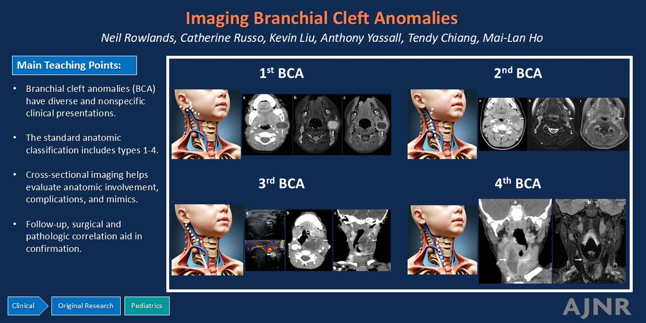
BACKGROUND AND PURPOSE: Branchial cleft anomalies (BCA) are a group of relatively common congenital pediatric neck masses, which exhibit wide variability in clinical presentation and treatment approaches. This study attempts to fill this gap by evaluating BCA clinical, radiographic, and treatment features in a large cross-sectional cohort.

MATERIALS AND METHODS: We performed a retrospective cross-sectional study of patients with BCA presenting to a single pediatric quaternary care center between 2017 and 2023. The radiology information system was queried for the term “branchial,” with diagnostic confirmation based on radiology and medical records review including patient demographics, clinical presentation and symptoms, genetic testing, interventions, and pathology. Relevant statistical tests were performed.

RESULTS: We retrospectively identified 302 unique patients with 412 imaging examinations that included the term “branchial” in the radiology information system between 2017 and 2023. The final cohort included 167 patients with a total of 246 imaging examinations. Among patients with BCA, median age at presentation was 3.3 years (range 0–22.9). Leading clinical presentations included a neck mass (88%, 147) and skin drainage (29%, 29). BCA classification was first in 37% (61), second in 44% (73), third in 4% (7), and fourth in 15% (26). Interventions included incision and drainage in 70% (121) and complete excision in 54% (91). Among patients with resected BCA, 22% (20/91) experienced at least 1 recurrence.

CONCLUSIONS: BCA have diverse clinical manifestations for which imaging aids in localization, classification, and interventional planning.

ABBREVIATIONS:

BCA
branchial cleft anomalies
FS
fat suppression
US
ultrasound

The branchial (pharyngeal) apparati are paired embryologic structures that mature during development into the anatomic structures of the head and neck. Occasionally, congenital remnants of the branchial clefts (ectoderm), arches (mesoderm), and/or pouches (endoderm) can persist into postnatal life. Branchial cleft anomalies (BCA) are common congenital pediatric neck masses and arise from persistent ectodermal remnants of the pharyngeal clefts. BCA are classified into 4 types depending on embryologic origin and extent of malformation (cyst, sinus, fistula).1,2

First BCA arise from the first branchial cleft involving the auricle, external auditory canal, parotid gland, and/or mandible. The Work classification is based on embryologic origin (type 1: postauricular with ectodermal elements, type 2: preauricular with ectodermal and mesodermal elements). The Arnot classification utilizes an anatomopathological classification (type 1: parotid gland with buried cell rests, type 2: anterior neck with communicating tract). The Olsen classification is based on communication with adjacent structures (cyst: closed, sinus: 1 opening, fistula: 2 openings) (Fig 1A).3

FIG 1.
  • Download figure
  • Open in new tab
  • Download powerpoint
FIG 1.

BCA classification based on embryologic tracts. A, First BCA is seen in the periauricular region or parotid gland. B, Second BCA involve the anterior triangle of neck such as submandibular triangle, sternocleidomastoid muscle, and carotid space. C, Third BCA can involve the posterior triangle or low anterior neck at the level of hypopharynx and piriform sinus base. D, Fourth BCA are located between the piriform sinus apex and thyroid gland in the visceral space.

Second BCA are the most common type and originate from the second branchial cleft, located within the anterior triangle of the neck. The Bailey classification subtypes lesions by anatomic location (type 1: superficial, between platysma and sternocleidomastoid muscle; type 2: anterior to sternocleidomastoid, posterior to submandibular gland, lateral to carotid sheath; type 3: medial carotid space between internal and external carotid arteries; type 4: parapharyngeal or pharyngeal mucosal space opening into lateral oropharynx) (Fig 1B).2,4

Third BCA arise from the third branchial cleft along the high posterior or low anterior neck, often involving the hypopharynx and piriform sinus base. The embryologic path lies near midline, below the glossopharyngeal nerve and above the superior laryngeal and glossopharyngeal nerves, although there is overlap with fourth BCA involving the recurrent laryngeal nerve (Fig 1C). Fourth BCA originate from the fourth branchial cleft and have anatomic overlap with third BCA, forming within the visceral space of the low anterior neck in the region of piriform sinus apex, thyroid gland, and mediastinum. Both third and fourth BCA are more common on the left than right, which may correspond with developmental resorption of the right fifth and sixth arches (Fig 1D).2,5,6

Clinical presentations of BCA are heterogeneous, ranging from asymptomatic to neck swelling or mass, pain, erythema, drainage, or recurrent suppurative thyroiditis with fourth BCA.1,2 Various imaging modalities can be used to characterize BCA location, composition, complications, and also exclude other potential etiologies. Ultrasound (US) enables visualization of superficial structures, differentiation of solid and cystic components, and evaluation of vascular structures via Doppler technique.7 For acute presentations, CT provides rapid cross-sectional evaluation with high spatial resolution, but involves ionizing radiation exposure with limited contrast resolution.5,8 MR imaging is the standard for anatomic characterization of complex soft tissue structures, however, may require sedation/anesthesia in younger children.9,10 Fluoroscopy can occasionally be useful for identifying fistulas while image-guided interventions can aid in minimally invasive percutaneous aspiration/biopsy for diagnosis or therapy.11

There are limited data regarding BCA clinical presentation, imaging characteristics, and surgical outcomes. This study was a comprehensive multidisciplinary review of branchial anomalies performed at a single quaternary children’s hospital to evaluate patient demographic characteristics, imaging distribution, associated genetic conditions, and interventional practices.

MATERIALS AND METHODS

The study was conducted with approval from the institutional review board at Nationwide Children’s Hospital in Columbus, Ohio. All data collection was conducted in compliance with the Health Insurance Portability and Accountability Act. The radiology information system was queried for the term “branchial” within the report and/or requisition of all imaging examinations performed between 2017 and 2023. All patients presenting to the freestanding children’s hospital were included, with 2 young adults (18 and 22 years) among them having medical histories necessitating care at a children’s hospital. All radiology examinations were initially interpreted by 1 of 6 fellowship-trained pediatric neuroradiologists with between 8 and 32 years of postgraduate experience, followed by confirmatory clinical assessment and follow-up by a board-certified pediatric otolaryngologist. Surgical or pathologic confirmation was obtained in 117 of 167 patients. A single fellowship-trained pediatric neuroradiologist with 10 years of postgraduate experience in pediatric head and neck imaging re-reviewed all imaging studies, surgical and pathologic findings, and medical records for adjudication of diagnosis. For patients with confirmed BCA, detailed information was collected from patient charts including age at presentation, sex, race, symptoms, imaging modalities, BCA laterality, BCA classification, genetic work-up, interventions, and recurrence after surgical excision. Imaging characteristics did not provide unique information beyond the currently established features of BCA and were not reported. Types of BCA were categorized by anatomic location. First BCA were periauricular or parotid (Fig 1), second in the anterior triangle of the neck (sternocleidomastoid muscle, submandibular gland, carotid spaces), third in the posterior triangle or low anterior neck at level of hypopharynx/base of piriform sinus, and fourth at the apex of the piriform sinus to the visceral space. Cases of cysts, sinuses, and fistulas were not recorded due to inconsistent visualization on imaging due to technique, small size/collapse, or obscuration by inflammation. These diagnoses are best identified by surgical exploration. Shapiro-Wilk and QQ plots were performed to determine if the continuous variables were parametric. Parametric variables were described in terms of mean and standard deviation, while nonparametric continuous variables were described in terms of median and interquartile range. Categoric and binary variables were reported as counts and proportions.

RESULTS

We retrospectively identified 302 patients with 412 imaging examinations that included the term “branchial” in the radiology requisition and/or report. After imaging and chart review, 166 examinations in 135 patients were excluded for alternate final diagnoses based on imaging, surgical, and/or pathologic information. Alternate diagnoses including lymphadenitis, skin pit/tag, dermoid/epidermoid cyst, and uncommon alternate diagnoses vascular malformation, duplication cyst, abscess, cellulitis, ranula/mucocele, thyroglossal duct cyst, and benign and malignant neoplasms, (lymphoma, neuroblastoma, nerve sheath tumor, thyroid adenoma, pilomatricoma, and keloid) were excluded. The final cohort included 167 patients with a confirmed diagnosis of BCA.

Of the 167 patients diagnosed with BCA, median age was 3.3 years (range 0 [neonate] to 22.9 years). Sex was 55% (92) girls and 45% (75) boys. Race was 65% (109) white, 20% (34) black, 5% (8) Latino, 3% (5) Asian, and 7% (11) multiracial.

Symptoms at time of presentation included acute infection 90% (150), neck mass or swelling 88% (147), skin drainage or pit/opening 29% (49), pain or tenderness to palpation 22% (36), skin erythema 14% (23), and neck stiffness or reduced range of motion 7% (11). Associated complications were airway-related (sore throat, voice changes, dysphonia, dysphagia) 11% (18), fever or other systemic symptoms 12% (20), and recurrence after initial symptom resolution 57% (95).

Underlying genetic conditions were suspected in 22% (36) of patients based on family history, abnormal facies, bilateral and/or multisystem findings. Genetic testing was positive for branchio-oto-renal syndrome in 23, Goldenhar syndrome in 7, and cardiofaciocutaneous syndrome in 1 patient (Table 1, Fig 2).

FIG 2.
  • Download figure
  • Open in new tab
  • Download powerpoint
FIG 2.

Branchio-oto-renal syndrome. A, Axial CT shows bilateral unwound cochleae (arrows) with tapered basal turns and hypoplastic offset middle/apical turns. B, Axial CT with contrast shows a first branchial cleft cyst (arrow) in the left external auditory canal. C, Balanced steady-state free precession MR shows hypoplastic cochleae and cochlear nerves (arrows).

DemographicsTypen (%)
Age3.3 (IQR 5.7–17.2)
SexBoy75 (45%)
Girl92 (55%)
RaceWhite109 (65%)
Black34 (20%)
Latino8 (5%)
Asian5 (3%)
Multiracial11 (7%)
SymptomsMass/swelling147 (88%)
Drainage/pit49 (29%)
Pain/tenderness36 (22%)
Erythema23 (14%)
Stiffness11 (7%)
Genetic syndromesBranchio-oto-renal23 (14%)
Goldenhar7 (4%)
Other6 (3%)
InterventionDrainage117 (70%)
Excision91 (54%)
Outcome after resectionRemission71 (78%)
Recurrence20 (22%)
  • Note:—IQR = interquartile range.

Table 1: Clinical characteristics for 167 patients with confirmed branchial cleft anomalies

A total of 246 imaging examinations were performed, with some patients receiving multiple imaging modalities. In the cohort of 167 patients, imaging included US 55% (135), CT 31% (77), MR 12% (29), and radiography/fluoroscopy 2% (5). Based on imaging findings with surgical or pathologic correlation, BCA laterality was to left in 49% (81), to right in 39% (65), and 13% (21) bilateral. Classification was 37% (61) first, 44% (73) second, 4% (7) third, and 15% (26) fourth (Table 2, Fig 3–6).

View this table:
  • View inline
  • View popup
Table 2:

Imaging modalities and BCA characteristics

FIG 3.
  • Download figure
  • Open in new tab
  • Download powerpoint
FIG 3.

First branchial cleft anomaly. A, Axial CT with contrast. B, T2-weighted FS MR. C, T1-weighted FS MR postcontrast shows a thick-walled rim-enhancing cystic lesion in the left parotid tail (arrows) with overlying inflammatory fat stranding and ipsilateral reactive lymphadenopathy.

FIG 4.
  • Download figure
  • Open in new tab
  • Download powerpoint
FIG 4.

Second branchial cleft anomaly. A, Axial CT with contrast shows a thin-walled rim-enhancing cystic lesion (arrow) in the left anterior neck between the submandibular gland, carotid space, and sternocleidomastoid. There is mild overlying fat stranding and ipsilateral lymphadenopathy. MR performed 1 month following antibiotic therapy with (B) T2-weighted FS and (C) T1-weighted FS postcontrast show interval decreased size of the collection (arrows) with resolution of fat stranding and lymphadenopathy.

FIG 5.
  • Download figure
  • Open in new tab
  • Download powerpoint
FIG 5.

Third branchial cleft anomaly. A, Transverse US with Doppler shows a complex cystic lesion with peripheral vascularity (arrows) superior to the left thyroid gland. CT with contrast, (B) axial, and (C) coronal images, show lobulated rim-enhancing cystic lesion contiguous with the base of left piriform sinus (arrows).

FIG 6.
  • Download figure
  • Open in new tab
  • Download powerpoint
FIG 6.

Fourth branchial cleft anomaly. A, Coronal CT with contrast shows a thick-walled rim-enhancing cystic lesion (arrow) within the right thyroid lobe, abutting the apex of pyriform sinus with overlying fat stranding and ipsilateral lymphadenopathy. After surgical cyst drainage and tract cautery, MR performed 1 year later with (B) T2-weighted FS shows lesion involution with faint residual edema (arrow) extending from the right piriform sinus apex to thyroid.

Interventions were required in most patients with 70% (117) undergoing incision and drainage, and 54% (91) proceeding to surgical excision. Surgical approaches varied based on BCA type, acuity, and surgeon and family preferences. Of patients who underwent surgical resection, 78% (71/91) were in full remission at 1-year follow-up, while 22% (20/91) experienced recurrence of clinical and/or imaging findings.

Pathology was sent for 112 patients and demonstrated features consistent with BCAs including respiratory epithelium with ciliated columnar cells, seromucinous contents with variable squamous metaplasia, and variable bulk fat with cartilage remnants. Secondary histologic findings included inflammation with granulation tissue; bacterial, viral, or granulomatous infection; and reactive lymph nodes.

DISCUSSION

BCA pose unique diagnostic and management challenges, with limited data regarding the diagnosis and follow-up of these pediatric anomalies. Multiple factors confound scientific analysis, including the diverse clinical presentations and mimics; heterogeneity of patient populations; propensity for recurrence with varying management approaches; inconsistent surgical techniques; and limited longitudinal follow-up. Therefore, references on this topic are limited to cross-sectional studies at large children’s hospitals, with the largest pediatric case series of 97 patients reported in 2007 over a 10-year period at a single hospital.11

Our large cohort represents a diverse spectrum of patient ages and demographics from neonate to young adult, with median age 3.3 years. The most common clinical presentation was a unilateral neck mass followed by drainage, pain, or erythema. Most patients were imaged by US and/or CT, reflecting the speed and ease of access of these modalities in the urgent setting. MR was utilized for deeper and complex lesions that presented nonemergently. In agreement with the literature, second BCA showed the highest incidence, followed by first BCA. Third/fourth BCA were rare and showed clinical overlap, requiring surgical evaluation of the superior laryngeal nerve for final classification. Twenty-two percent of patients with BCA had associated genetic conditions, particularly when findings were bilateral (most commonly first BCA in branchio-oto-renal syndrome). More than one-half of patients experienced recurrence of symptoms after initial resolution, requiring surgical drainage and/or total excision. Overall, our results indicate the need for a multidisciplinary approach to BCA diagnosis and management.

Postoperative outcomes are highly dependent on BCA type and complexity, as well as surgical technique, which varies greatly between centers. When the BCA and tract are completely excised, recurrence rates are low at 2%–4%.12,14 However, complete resection often involves extensive and invasive surgery. Many centers are moving toward minimally invasive endoscopic approaches with shorter patient recovery times. With this approach, there is an increased risk of incomplete excision, in which case recurrence rates can reach 10%–20% or more.15⇓⇓⇓–19 In our study, 57% of patients experienced at least 1 recurrence after medical management or drainage, compared with only 22% of patients after surgical excision. Patients who had recurrence after surgery tended to have more complex anatomy, including branchio-oto-renal syndrome cases with bilateral first BCA, and third/fourth BCA with persistent airway and/or thyroid communications. These patients required additional interventions and follow-up with otolaryngology providers to achieve permanent resolution.

Our study was inherently limited by a single reviewer determining inclusion and BCA classification, and the retrospective single-center design and requirement of imaging for inclusion. Our multidisciplinary evaluation was critical for confirming BCA diagnosis and assessing follow-up results, but precludes separate quantification of imaging, surgical, and pathologic accuracy in BCA. Ultimately, multiple aspects of radiologic, surgical, and pathologic technique are highly individualized and depend greatly on patient presentation and physician experience. Nevertheless, our data represent the newest and largest known imaging case series of BCA and provide novel insights into clinical characteristics, lesion classification, and management.

CONCLUSIONS

BCA have diverse clinical presentations and the tendency to recur, generating multiple diagnostic and management challenges. Cross-sectional imaging is important to confirm the diagnosis and establish location, laterality, and extent of BCA for interventional planning. BCA classification influences the surgical approach and outcome, with multidisciplinary collaboration important to achieve optimal patient outcomes. Continued evaluation and standardization of diagnostic workflow and management approaches across centers will be key to future research endeavors.

Footnotes

  • Disclosure forms provided by the authors are available with the full text and PDF of this article at www.ajnr.org.

References

  1. 1.↵
    1. Mehmi N,
    2. Kumar R,
    3. Sagar P, et al
    . Importance and impact of appropriate radiology in the management of branchial cleft anomalies. Indian J Otolaryngol Head Neck Surg 2019;71:953–59 doi:10.1007/s12070-019-01634-w pmid:31742101
    CrossRefPubMed
  2. 2.↵
    1. Adams A,
    2. Mankad K,
    3. Offiah C, et al
    . Branchial cleft anomalies: a pictorial review of embryological development and spectrum of imaging findings. Insights Imaging 2016;7:69–76 doi:10.1007/s13244-015-0454-5 pmid:26661849
    CrossRefPubMed
  3. 3.↵
    1. D’Souza AR,
    2. Uppal HS,
    3. De R, et al
    . Updating concepts of first branchial cleft defects: a literature review. Int J Pediatr Otorhinolaryngol 2002;62:103–09 doi:10.1016/s0165-5876(01)00612-7 pmid:11788142
    CrossRefPubMed
  4. 4.↵
    1. Bajaj Y,
    2. Ifeacho S,
    3. Tweedie D, et al
    . Branchial anomalies in children. Int J Pediatr Otorhinolaryngol 2011;75:1020–23 doi:10.1016/j.ijporl.2011.05.008 pmid:21680029
    CrossRefPubMed
  5. 5.↵
    1. Thomas B,
    2. Shroff M,
    3. Forte V, et al
    . Revisiting imaging features and the embryologic basis of third and fourth branchial anomalies. AJNR Am J Neuroradiol 2010;31:755–60 doi:10.3174/ajnr.A1902 pmid:20007720
    Abstract/FREE Full Text
  6. 6.↵
    1. Waldhausen JH
    . Branchial cleft and arch anomalies in children. Semin Pediatr Surg 2006;15:64–69 doi:10.1053/j.sempedsurg.2006.02.002 pmid:16616308
    CrossRefPubMed
  7. 7.↵
    1. Bansal AG,
    2. Oudsema R,
    3. Masseaux JA, et al
    . US of pediatric superficial masses of the head and neck. Radiographics 2018;38:1239–63 doi:10.1148/rg.2018170165 pmid:29995618
    CrossRefPubMed
  8. 8.↵
    1. Harnsberger HR,
    2. Mancuso AA,
    3. Muraki AS, et al
    . Branchial cleft anomalies and their mimics: computed tomographic evaluation. Radiology 1984;152:739–48 doi:10.1148/radiology.152.3.6463255 pmid:6463255
    CrossRefPubMed
  9. 9.↵
    1. Black CJ,
    2. O’Hara JT,
    3. Berry J, et al
    . Magnetic resonance imaging of branchial cleft abnormalities: illustrated cases and literature review. J Laryngol Otol 2010;124:213–15 doi:10.1017/S0022215109990995 pmid:19765327
    CrossRefPubMed
  10. 10.↵
    1. Unsal O,
    2. Soytas P,
    3. Hascicek SO, et al
    . Clinical approach to pediatric neck masses: retrospective analysis of 98 cases. North Clin Istanb 2017;4:225–32 doi:10.14744/nci.2017.15013 pmid:29270570
    CrossRefPubMed
  11. 11.↵
    1. Schroeder JW, Jr.,
    2. Mohyuddin N,
    3. Maddalozzo J
    . Branchial anomalies in the pediatric population. Otolaryngol Head Neck Surg 2007;137:289–95 doi:10.1016/j.otohns.2007.03.009 pmid:17666258
    CrossRefPubMed
  12. 12.↵
    1. Mattioni J,
    2. Azari S,
    3. Hoover T, et al
    . A cross-sectional evaluation of outcomes of pediatric branchial cleft cyst excision. Int J Pediatr Otorhinolaryngol 2019;119:171–76 doi:10.1016/j.ijporl.2019.01.030 pmid:30735909
    CrossRefPubMed
  13. 13.
    1. Raghavan M,
    2. Carr MM
    . Age-related outcomes after pediatric branchial cleft cyst excision via NSQIP-P. Int J Pediatr Otorhinolaryngol 2024;176:111811 doi:10.1016/j.ijporl.2023.111811 pmid:38048733
    CrossRefPubMed
  14. 14.↵
    1. Ford GR,
    2. Balakrishnan A,
    3. Evans JN, et al
    . Branchial cleft and pouch anomalies. J Laryngol Otol 1992;106:137–43 doi:10.1017/s0022215100118900 pmid:1556487
    CrossRefPubMed
  15. 15.↵
    1. Al-Mufarrej F,
    2. Stoddard D,
    3. Bite U
    . Branchial arch anomalies: recurrence, malignant degeneration and operative complications. Int J Pediatr Otorhinolaryngol 2017;97:24–29 doi:10.1016/j.ijporl.2017.03.014 pmid:28483243
    CrossRefPubMed
  16. 16.↵
    1. Quintanilla-Dieck L,
    2. Virgin F,
    3. Wootten C, et al
    . Surgical approaches to first branchial cleft anomaly excision: a case series. Case Rep Otolaryngol 2016;2016:3902974 doi:10.1155/2016/3902974 pmid:27034873
    CrossRefPubMed
  17. 17.↵
    1. Paul I,
    2. Mohiyuddin SMA,
    3. Sagayaraj A, et al
    . The outcome of treatment in second branchial cleft anomalies: a case series. Cureus 2023;15:e40164 doi:10.7759/cureus.40164 pmid:37431352
    CrossRefPubMed
  18. 18.↵
    1. Teng SE,
    2. Paul BC,
    3. Brumm JD, et al
    . Endoscope-assisted approach to excision of branchial cleft cysts. Laryngoscope 2016;126:1339–42 doi:10.1002/lary.25711 pmid:26466762
    CrossRefPubMed
  19. 19.↵
    1. Derks LS,
    2. Veenstra HJ,
    3. Oomen KP, et al
    . Surgery versus endoscopic cauterization in patients with third or fourth branchial pouch sinuses: a systematic review. Laryngoscope 2016;126:212–17 doi:10.1002/lary.25321 pmid:26372400
    CrossRefPubMed
  • Received July 15, 2024.
  • Accepted after revision October 9, 2024.
  • © 2025 by American Journal of Neuroradiology
PreviousNext
Back to top
Advertisement
Print
Download PDF
Email Article

Thank you for your interest in spreading the word on American Journal of Neuroradiology.

NOTE: We only request your email address so that the person you are recommending the page to knows that you wanted them to see it, and that it is not junk mail. We do not capture any email address.

Enter multiple addresses on separate lines or separate them with commas.
Clinical Characteristics of Branchial Cleft Anomalies
(Your Name) has sent you a message from American Journal of Neuroradiology
(Your Name) thought you would like to see the American Journal of Neuroradiology web site.
CAPTCHA
This question is for testing whether or not you are a human visitor and to prevent automated spam submissions.
Cite this article
Neil Rowlands, Catherine Russo, Kevin Liu, Anthony Yassall, Tendy Chiang, Mai-Lan Ho
Clinical Characteristics of Branchial Cleft Anomalies
American Journal of Neuroradiology Mar 2025, DOI: 10.3174/ajnr.A8604

Citation Manager Formats

  • BibTeX
  • Bookends
  • EasyBib
  • EndNote (tagged)
  • EndNote 8 (xml)
  • Medlars
  • Mendeley
  • Papers
  • RefWorks Tagged
  • Ref Manager
  • RIS
  • Zotero
0 Responses
Respond to this article
Share
Bookmark this article
Imaging Branchial Cleft Anomalies
Neil Rowlands, Catherine Russo, Kevin Liu, Anthony Yassall, Tendy Chiang, Mai-Lan Ho
American Journal of Neuroradiology Mar 2025, DOI: 10.3174/ajnr.A8604
del.icio.us logo Twitter logo Facebook logo Mendeley logo
  • Tweet Widget
  • Facebook Like
  • Google Plus One
Purchase

Jump to section

  • Article
    • Graphical Abstract
    • Abstract
    • ABBREVIATIONS:
    • MATERIALS AND METHODS
    • RESULTS
    • DISCUSSION
    • CONCLUSIONS
    • Footnotes
    • References
  • Figures & Data
  • Info & Metrics
  • Responses
  • References
  • PDF

Related Articles

  • No related articles found.
  • PubMed
  • Google Scholar

Cited By...

  • No citing articles found.
  • Crossref
  • Google Scholar

This article has not yet been cited by articles in journals that are participating in Crossref Cited-by Linking.

More in this TOC Section

  • Frontal Paraventricular Cysts
  • Sodium MRI in Pediatric Brain Tumors
  • FRACTURE MR in Congenital Vertebral Anomalies
Show more Pediatric Neuroimaging

Similar Articles

Advertisement

Indexed Content

  • Current Issue
  • Accepted Manuscripts
  • Article Preview
  • Past Issues
  • Editorials
  • Editor's Choice
  • Fellows' Journal Club
  • Letters to the Editor
  • Video Articles

Cases

  • Case Collection
  • Archive - Case of the Week
  • Archive - Case of the Month
  • Archive - Classic Case

More from AJNR

  • Trainee Corner
  • Imaging Protocols
  • MRI Safety Corner
  • Book Reviews

Multimedia

  • AJNR Podcasts
  • AJNR Scantastics

Resources

  • Turnaround Time
  • Submit a Manuscript
  • Submit a Video Article
  • Submit an eLetter to the Editor/Response
  • Manuscript Submission Guidelines
  • Statistical Tips
  • Fast Publishing of Accepted Manuscripts
  • Graphical Abstract Preparation
  • Imaging Protocol Submission
  • Evidence-Based Medicine Level Guide
  • Publishing Checklists
  • Author Policies
  • Become a Reviewer/Academy of Reviewers
  • News and Updates

About Us

  • About AJNR
  • Editorial Board
  • Editorial Board Alumni
  • Alerts
  • Permissions
  • Not an AJNR Subscriber? Join Now
  • Advertise with Us
  • Librarian Resources
  • Feedback
  • Terms and Conditions
  • AJNR Editorial Board Alumni

American Society of Neuroradiology

  • Not an ASNR Member? Join Now

© 2025 by the American Society of Neuroradiology All rights, including for text and data mining, AI training, and similar technologies, are reserved.
Print ISSN: 0195-6108 Online ISSN: 1936-959X

Powered by HighWire