Abstract
BACKGROUND AND PURPOSE: Patients with intracranial hypotension from spinal CSF leaks have increased choroid plexus volumes in response to CSF leakage. The purpose of this study was to assess changes in choroid plexus volumes in patients before and after spinal CSF leak repair.
MATERIALS AND METHODS: This was a retrospective, institutional review board–approved study on patients with spinal CSF leak who had pre- and post-CSF leak repair MRI examinations. Brain MRIs with contrast were performed on a 1.5/3T scanner with acquisition of 3D T1 postcontrast (eg, Bravo, MPRAGE, and so forth). Choroid plexus volumes at the level of the trigonum ventriculi were calculated for the left and right sides on all pre- and posttreatment MRIs using Visage-7 segmentation tools. Basic demographic data, type of CSF leak, and choroid plexus volumes were recorded for all patients. Basic 2-tailed t tests were used to compare choroid plexus volumes between the pre- and posttreatment groups.
RESULTS: Twenty patients with spontaneous intracranial hypotension from spinal CSF leaks were included. Eleven patients (55%) had a type 1a (ventral tear) spinal CSF leak, 5 patients (25%) had type 1b (lateral tear), and 4 patients (20%) had a type 3 spinal CSF leak. The mean age was 47.6 years (SD, 13.8 years). The mean choroid plexus volumes pretreatment were 0.82 cm3 (SD, 0.29 cm3) compared with 0.38 cm3 (SD, 0.19 cm3) posttreatment (P value 0.01).
CONCLUSIONS: Significantly decreased choroid plexus volumes were seen in patients with spontaneous intracranial hypotension following spinal CSF leak repair. This finding highlights the modulation and dynamic role of the choroid plexus in states of low CSF volumes.
ABBREVIATION:
- SIH
- spontaneous intracranial hypotension
The choroid plexus comprises specialized cells charged with modulating CSF production.1⇓-3 Prior studies have shown that the choroid plexus is a dynamic structure not only demonstrating changes during the life span but also playing a role in neuroinflammation and neuroimmune modulation.4⇓⇓⇓⇓⇓⇓⇓⇓⇓⇓-15 Additionally, the modulation of CSF volume is also an important role of the choroid plexus, with prior studies demonstrating hyperplasia of the choroid plexus in the setting of hydrocephalus.16,17 A recent study also demonstrated increased volumes of the choroid plexus in patients with spinal CSF leaks compared with age- and sex-matched healthy controls.18 This work reveals that volumetric changes of the choroid plexus occur in states of low CSF volume via compensatory modulation. Potentially, choroid plexus volumes may be used as an independent biomarker for CSF homeostasis and may potentially be added to the criteria assessing spontaneous intracranial hypotension (SIH) such as the Bern score; however, additional investigations into this topic are needed.19⇓⇓-22 The purpose of this study was to evaluate intrapatient changes in choroid plexus volumes for individuals with spinal CSF leak before and after successful leak repair.
MATERIALS AND METHODS
This was a retrospective, institutional review board–approved study performed at a single institution. Patients were included in this study if they met the following conditions: 1) adult patients, 2) with clinical and imaging features of SIH on brain MRI, 3) who had a spinal CSF leak diagnosed on CT myelography, 4) who underwent surgical repair of the spinal CSF leak at our institution, and 5) who had a post-CSF leak treatment MRI. Patients were excluded from this cohort if they had MRI examinations that were severely degraded by artifacts and were not of diagnostic quality. Additionally, patients who underwent CSF leak treatment but achieved a suboptimal response (defined as incomplete symptom resolution, persistence of symptoms, and/or lack of resolution of intracranial imaging features of SIH on MRI) were excluded.
All brain MRI examinations in this study were performed with gadolinium contrast and included an isotropic 3D T1 sequence (such as Bravo [GE Healthcare], MPRAGE, and so forth). Pretreatment MRI brain studies were evaluated over a 7-year time period from 2017 to 2023 and were all performed immediately before identification of the spinal CSF leak on myelography. Posttreatment MRI brain studies were performed after surgical repair during an 8-year period from 2017 to 2024. Examinations were acquired across multiple MRI vendor platforms and included 1.5T and 3T magnet strengths (Signa Artist, Signa HX, and Signa Excite HDx; GE Healthcare; Magnetom Prisma Fit and Magnetom Skyra; Siemens).
Manual 3D T1-postcontrast choroid plexus contouring was performed in Visage-7 (Visage Imaging) using the 3D Freehand tool ROI under the segmentation toolkit (Visage, Version 7.1.18). From these manual contours, a 3D volume of the choroid plexus was calculated. This was performed for both the left and right sides at the trigonum ventriculi, using a technique previously published in the literature and shown in Fig 1.18,23-24 The choroid plexus volumes were independently calculated for the left and right sides in all patients on both the pretreatment and posttreatment scans and were counted as 4 independent and discrete data points (ie, 2 choroid plexus volumes on pretreatment MRI and 2 choroid plexus volumes on posttreatment MRI per patient). Using the methodology previously published, if present, we excluded choroid plexus cysts and xanthogranuloma from the volumetric contours.18 Choroid plexus contours were performed by 2 neuroradiologists with Certificates of Added Qualification.

A 65-year-old woman with a type 3 CSF leak, status post surgical repair presenting for routine follow-up brain MRI. Axial T1 postcontrast MPRAGE sequences are shown demonstrating a manual segmentation of the left choroid plexus. A magnified view of the same patient; the same slice is shown on the right-hand panel.
Basic demographic data including patient age, sex, type of spinal CSF leak, pretreatment, and Bern score were recorded for all patients.22,25
Statistical analysis using a 2-tailed t test was used to compare choroid plexus volumes between pretreatment and posttreatment MRI examinations for the patients with SIH with spinal CSF leaks. Additionally, a correlation coefficient was calculated between the mean of the left and right choroid plexus volumes in each patient compared with the pretreatment Bern score. The methodology proposed in the Strengthening the Reporting of Observational Studies in Epidemiology (STROBE) checklist was followed.
RESULTS
Twenty patients were included in this study, with a total of 80 recorded choroid plexus volumes (2 pretreatment volumes and 2 posttreatment volumes for each patient). The mean patient age was 47.6 years (SD, 13.8 years; range, 25–65 years). There were 8 men and 12 women in this cohort.
There were 11 patients (55%) with type 1a (ventral tear) spinal CSF leak, 5 patients with type 1b (lateral tear) leaks, and 4 (20%) patients with a type 3 spinal CSF leak. Pretreatment MRI scans all demonstrated imaging features consistent with SIH with a mean Bern score of 7 (SD, 1.6; range, 5–9). Posttreatment MRI scans demonstrated complete resolution of intracranial imaging features of SIH and a Bern score of zero for all patients.
The mean choroid plexus volume on pretreatment examinations was 0.82 cm3 (SD, 0.3; interquartile range, 0.33), compared to posttreatment mean volumes of 0.38 cm3 (SD, 0.22; interquartile range, 0.28) (P value < .0001) (Fig 2). A summary of this demographic, clinical, and imaging data is shown in the Table, and a representative case is shown in Fig 2. There was no statistically significant correlation between the pretreatment Bern score and the mean choroid plexus volume for each patient (r = 0.02, P = .93). For the patients with types 1a and 1b spinal CSF leaks, the mean choroid plexus pretreatment volume was 0.8 cm3 with a mean choroid plexus posttreatment volume of 0.4 cm3, a 50% reduction. For the type 3 spinal CSF leaks, the mean pretreatment choroid plexus volume was 0.9 cm3 compared with 0.35 cm3 posttreatment, a 38% reduction.

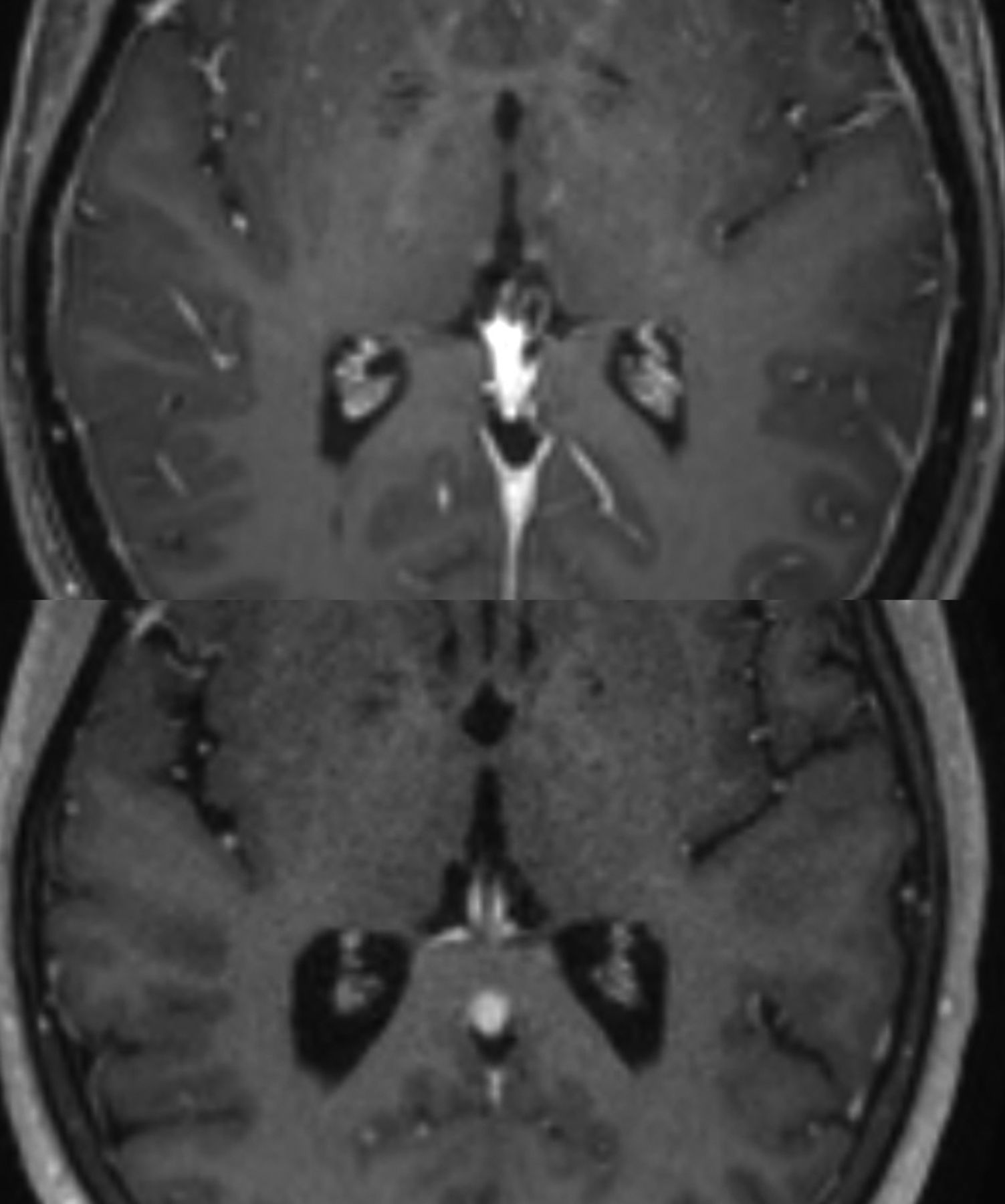
Axial T1 postcontrast MPRAGE sequences at the level of the atria in the same 65-year-old woman with a type 3 spinal CSF leak shown in Fig 1. The upper image demonstrates the pretreatment brain MRI. The lower image shows the posttreatment brain MRI. By visual inspection, the volume of the choroid plexus on the pretreatment examination compared with the posttreatment MRI has markedly decreased.
The time between the pretreatment brain MRI and posttreatment brain MRI was somewhat variable with a mean follow-up interval of 25 months (range, 1–76 months).
DISCUSSION
A statistically significant decrease in the volumes of the choroid plexus was seen on the posttreatment MRI scans compared with pretreatment MRI scans for patients with spinal CSF leaks. In this group of patients who had successful treatment of known spinal CSF leaks, defined as complete resolution of symptoms and imaging findings suggestive of SIH, the choroid plexus demonstrates volumetric modulation based on changing states of CSF volumes. Prior research has shown that choroid plexus volumes in patients with spinal CSF leaks are significantly larger than those in healthy controls,18 this study builds off of that prior research suggesting that the enlargement of the choroid plexus may be transient and reversible if CSF leakage can be successfully stopped. This finding is thought to demonstrate the dynamic and compensatory ability of the choroid plexus to downregulate CSF production once the spinal CSF leak is treated.1,2,26,27 As postulated previously, both hyperplasia and vascular engorgement related to expansion of the intracranial blood pool in SIH may have contributed to higher pretreatment choroid plexus volumes in these patients. It is also possible that the compensatory increases in CSF production in patients with spinal CSF leak could be via other mechanisms, including the upregulation of transporter proteins in epithelial cells by the choroid plexus and changes in blood volume to the choroid stroma.28⇓⇓-31 There may be downregulation of choroid plexus membrane proteins as well as decreased perfusion and blood pool following successful CSF leak repair. Further work and investigations into the mechanisms of modulation by the choroid plexus in patients with spinal CSF leaks are needed.
This study has limitations. First, the sample size is relatively small. While spinal CSF leaks are becoming an increasingly recognized pathology, their overall incidence remains relatively low. Furthermore, posttreatment MRI scanning was variable in our spinal CSF study population, likely reflective of our institution being a tertiary referral center for the treatment of spinal CSF leaks, attracting patients from across the nation, as well as heterogeneity within our own providers with respect to standardized posttreatment follow-up imaging. The lack of a standardized diagnostic work-up and posttreatment imaging and assessment of these patients with spinal CSF leak results in data heterogeneity, reduces cohort numbers, and limits the ability for multi-institutional comparisons/studies. This sentiment has been previously echoed by others in the spinal CSF leak literature.32,33 As a result, the time between spinal CSF leak repair and posttreatment MRI was variable. Additionally, this study looked solely at choroid plexus volumes as the primary metric. For future direction, adding a functional component to the structural, volumetric component would help gain additional insights into the dynamic and modulating function of the choroid plexus.
Furthermore, potential confounders such as medication usage, body mass index, blood pressure, and hydration status were not considered in this study and may impact the generalizability of these results. Last, patients were scanned on a combination of 1T and 3T platforms. Some differences in scanner strengths on pretreatment and posttreatment imaging may contribute to potential differences in choroid plexus volumes.
CONCLUSIONS
Choroid plexus volumes significantly decreased in spinal CSF leaks following repair of the leaks compared with their pretreatment volumes. This finding reflects the dynamic nature of the choroid plexus likely aiding in the modulation of CSF production in SIH. Future investigations into these structural changes in various CSF volumetric states may aid in the determination of choroid plexus volumes as a potential biomarker.
| Pretreatment Patients with SIH (n = 20) | Posttreatment Patients with SIH (n = 20) | P Value | |
|---|---|---|---|
| Age (mean, SD) (yr) | 47.6, 13.8 | ||
| Sex (M/F) | 8:12 | ||
| Bern score (mean, SD) | 7.1, 1.6 | 0, 0 | <.0001 |
| Type of spinal CSF leak | |||
| Type 1a | 11 | ||
| Type 1b | 5 | ||
| Type 3 | 4 | ||
| CP volume (mean, SD) (cm3) | 0.82, 0.29 | 0.38, 0.22 | <.0001 |
Note:—CP indicates choroid plexus; M, male; F, female.
a Pretreatment spinal CSF choroid plexus volumes were compared with posttreatment spinal CSF choroid plexus volumes for a cohort of 20 patients.
Basic demographic, clinical, and imaging features of patients with SIHa
Footnotes
Disclosure forms provided by the authors are available with the full text and PDF of this article at www.ajnr.org.
References
- 1.
- 2.
- 3.
- 4.
- 5.
- 6.
- 7.
- 8.
- 9.
- 10.
- 11.
- 12.
- 13.
- 14.
- 15.
- 16.
- 17.
- 18.
- 19.
- 20.
- 21.
- 22.
- 23.
- 24.
- 25.
- 26.
- 27.
- 28.
- 29.
- 30.
- 31.
- 32.
- 33.
- Received August 3, 2024.
- Accepted after revision August 28, 2024.
- © 2025 by American Journal of Neuroradiology