Advertisement

Main menu

  • Home
  • Content
    • Current Issue
    • Accepted Manuscripts
    • Article Preview
    • Past Issue Archive
    • Video Articles
    • AJNR Case Collection
    • Case of the Week Archive
    • Case of the Month Archive
    • Classic Case Archive
  • Special Collections
    • AJNR Awards
    • Low-Field MRI
    • Alzheimer Disease
    • ASNR Foundation Special Collection
    • Photon-Counting CT
    • View All
  • Multimedia
    • AJNR Podcasts
    • AJNR SCANtastic
    • Trainee Corner
    • MRI Safety Corner
    • Imaging Protocols
  • For Authors
    • Submit a Manuscript
    • Submit a Video Article
    • Submit an eLetter to the Editor/Response
    • Manuscript Submission Guidelines
    • Statistical Tips
    • Fast Publishing of Accepted Manuscripts
    • Graphical Abstract Preparation
    • Imaging Protocol Submission
    • Author Policies
  • About Us
    • About AJNR
    • Editorial Board
    • Editorial Board Alumni
  • More
    • Become a Reviewer/Academy of Reviewers
    • Subscribers
    • Permissions
    • Alerts
    • Feedback
    • Advertisers
    • ASNR Home

User menu

  • Alerts
  • Log in

Search

  • Advanced search
American Journal of Neuroradiology

American Journal of Neuroradiology

ASHNR American Society of Functional Neuroradiology ASHNR American Society of Pediatric Neuroradiology ASSR
  • Alerts
  • Log in

Advanced Search

  • Home
  • Content
    • Current Issue
    • Accepted Manuscripts
    • Article Preview
    • Past Issue Archive
    • Video Articles
    • AJNR Case Collection
    • Case of the Week Archive
    • Case of the Month Archive
    • Classic Case Archive
  • Special Collections
    • AJNR Awards
    • Low-Field MRI
    • Alzheimer Disease
    • ASNR Foundation Special Collection
    • Photon-Counting CT
    • View All
  • Multimedia
    • AJNR Podcasts
    • AJNR SCANtastic
    • Trainee Corner
    • MRI Safety Corner
    • Imaging Protocols
  • For Authors
    • Submit a Manuscript
    • Submit a Video Article
    • Submit an eLetter to the Editor/Response
    • Manuscript Submission Guidelines
    • Statistical Tips
    • Fast Publishing of Accepted Manuscripts
    • Graphical Abstract Preparation
    • Imaging Protocol Submission
    • Author Policies
  • About Us
    • About AJNR
    • Editorial Board
    • Editorial Board Alumni
  • More
    • Become a Reviewer/Academy of Reviewers
    • Subscribers
    • Permissions
    • Alerts
    • Feedback
    • Advertisers
    • ASNR Home
  • Follow AJNR on Twitter
  • Visit AJNR on Facebook
  • Follow AJNR on Instagram
  • Join AJNR on LinkedIn
  • RSS Feeds

AJNR Awards, New Junior Editors, and more. Read the latest AJNR updates

Sturge-Weber Syndrome

  • Background:
    • Rare, congenital, sporadic, neurocutaneous, and vascular disorder caused by somatic mutations in fetal ectodermal tissues that cause dysregulation of capillary blood vessel formation
    • Angiomas of the face, leptomeninges, and choroid are responsible for many of the signs and symptoms of this condition.
    • Many of the neurologic sequelae are thought to result from 2 pathophysiologic mechanisms—leptomeningeal capillary venous malformation and venous congestion—that both contribute to hypoxic-ischemic tissue injury.
  • Clinical Presentation:
    • Port-wine stains of the upper eyelid and forehead and other areas innervated by the trigeminal nerve (facial capillary malformation) are the most common type of vascular malformation; seizures, focal neurologic defects, intellectual disability, visual field defects, and glaucoma are also seen.
  • Key Diagnostic Features:
    • MRI T1/T2 with gadolinium: Leptomeningeal enhancement from congested cerebral veins and consequent parenchymal ischemia. Early phase changes may include restricted diffusion in an acute ischemic event. Late phase changes can demonstrate chronic ischemia and resultant gliosis with increased signal. Gyriform calcifications and enlarged transmedullary veins.
    • CT: Subcortical calcification and parenchymal volume loss; prominent choroid plexus
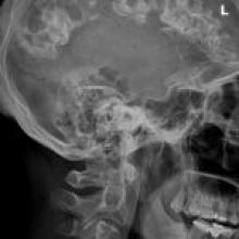
    • X-ray: Gyriform calcification of subcortical white matter known as “tram-track sign”
  • Differential Diagnoses:
    • Cerebral AVM: Hyperdense nidus of blood density on noncontrast CT and a “bag of worms” appearance on CT with contrast
    • Developmental venous anomaly (DVA): Enlarged draining veins and dystrophic calcification on noncontrast CT; linear or curvilinear focus of enhancement, typically coursing from the deep white matter to a cortical or a deep vein or to the dural sinus
    • Gobbi syndrome, also known as CEC syndrome (celiac disease, epilepsy, cerebral calcifications): Rarely present with bilateral occipital calcifications secondary to chronic venous ischemia on CT (absence of lobar or hemispheric atrophy or contrast enhancement, all of which may be present in Sturge-Weber)
    • Cerebral proliferative angiopathy: Tubular and linear parenchymal and subcortical hyperdensities on noncontrast CT; tubular flow voids on T1-weighted and T2-weighted sequences with abnormal, enlarged serpiginous vessels seen on contrast-enhanced images; multiple small arterial feeders and numerous small draining veins with no dominant feeders or nidus; diffuse or proliferative-type vascular malformation with normal cerebral tissue in between the abnormal vessels (though chronic hemorrhage, gliosis, or both can be seen in the brain tissue adjacent to the abnormal vessels); dilated proximal intracranial or extracranial arteries may also be demonstrated.
    • Meningioangiomatosis: Mainly cortical but also subcortical lesions of nodular or gyriform configuration; more focal than Sturge-Weber and typically lacks associated atrophy; demonstrates meningovascular proliferation and calcification and so manifests with enhancement; nonspecific imaging findings; on CT - low density, contrast enhancement, and intralesional calcifications; on MRI - hypointensity on T1 and hyperintensity on T2; spares the white matter; may be interpreted as a low-grade glioma
  • Treatment:
    • No specific treatment exists.
    • Anticonvulsant therapy has shown mixed results for treating seizures.
    • Surgery for refractory seizures
    • Port-wine stains can be treated with photothermolysis laser treatment.
    • Glaucoma is most effectively treated surgically.
December 23, 2021

A 45-year-old man with neck pain

View Case
  • Read more about 12232021
  • Comments
Advertisement
Subscribe to RSS - Sturge-Weber Syndrome

Indexed Content

  • Current Issue
  • Accepted Manuscripts
  • Article Preview
  • Past Issues
  • Editorials
  • Editor's Choice
  • Fellows' Journal Club
  • Letters to the Editor
  • Video Articles

Cases

  • Case Collection
  • Archive - Case of the Week
  • Archive - Case of the Month
  • Archive - Classic Case

More from AJNR

  • Trainee Corner
  • Imaging Protocols
  • MRI Safety Corner
  • Book Reviews

Multimedia

  • AJNR Podcasts
  • AJNR Scantastics

Resources

  • Turnaround Time
  • Submit a Manuscript
  • Submit a Video Article
  • Submit an eLetter to the Editor/Response
  • Manuscript Submission Guidelines
  • Statistical Tips
  • Fast Publishing of Accepted Manuscripts
  • Graphical Abstract Preparation
  • Imaging Protocol Submission
  • Evidence-Based Medicine Level Guide
  • Publishing Checklists
  • Author Policies
  • Become a Reviewer/Academy of Reviewers
  • News and Updates

About Us

  • About AJNR
  • Editorial Board
  • Editorial Board Alumni
  • Alerts
  • Permissions
  • Not an AJNR Subscriber? Join Now
  • Advertise with Us
  • Librarian Resources
  • Feedback
  • Terms and Conditions
  • AJNR Editorial Board Alumni

American Society of Neuroradiology

  • Not an ASNR Member? Join Now

© 2025 by the American Society of Neuroradiology All rights, including for text and data mining, AI training, and similar technologies, are reserved.
Print ISSN: 0195-6108 Online ISSN: 1936-959X

Powered by HighWire