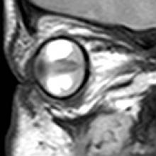
Persistent Hyperplastic Primary Vitreous (PHPV)
- During early gestation, primary vitreous, a normal embryonic hyaloid vasculature supplies nutrients to the developing lens and retina. This should regress completely by the 8th month of gestation, failure of which results in retrolental masses known as PHPV. Two types of PHPV: anterior and posterior.
- Associations: fetal alcohol syndrome, fetal hydantoin syndrome, and midline congenital cranial defects
- 90% cases are unilateral. Bilateral cases associated with trisomies 13, 15, 18, and 21.
- Children often present with leukocoria and microphthalmia.
- Imaging is necessary to rule out retinoblastoma.
- Key Diagnostic Features: Microphthalmia, shallow anterior chamber, abnormal density, and signal intensity consistent with hemorrhage in the vitreous chamber, retinal detachment, and enhancing retrolental/intravitreal tissue. Cloquet's canal can be seen. Calcification is absent. Associated findings: malformations of optic nerve and retina.
- DDx: Retinoblastoma, retinopathy of prematurity
- Rx: Lensectomy, removal of PHPV, and creation of black pupil (cosmetic)