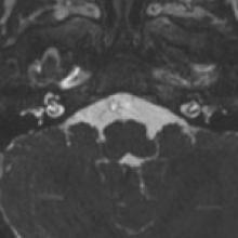
Labyrinthitis Ossificans (Fibrous Stage)
- Labyrinthitis ossificans refers to ossification of spaces within the lumen of the bony labyrinth and cochlea. It most commonly occurs as a sequela of inflammation of the inner ear that results from bacterial meningitis and subsequent purulent labyrinthitis.
- There are three characteristic stages in the process: acute, fibrous, and ossifying.
- Patient evaluation can be performed with CT or MRI, however, fibrosis may not always be obvious on CT. On MRI, the bright fluid of the normal cochlea is absent on the affected side. Although this may seen in FSE T2 images, it is better depicted on CISS or FIESTA high- resolution T2 sequences.
- CT detects ossification within the cochlea during the later stage of the process; it may not detect early ossification. The diagnosis of fibrosis is very important if cochlear implantation is considered as it may prevent or cause difficulties in insertion of electrode.


