Advertisement

Main menu

  • Home
  • Content
    • Current Issue
    • Accepted Manuscripts
    • Article Preview
    • Past Issue Archive
    • Video Articles
    • AJNR Case Collection
    • Case of the Week Archive
    • Case of the Month Archive
    • Classic Case Archive
  • Special Collections
    • AJNR Awards
    • Low-Field MRI
    • Alzheimer Disease
    • ASNR Foundation Special Collection
    • Photon-Counting CT
    • View All
  • Multimedia
    • AJNR Podcasts
    • AJNR SCANtastic
    • Trainee Corner
    • MRI Safety Corner
    • Imaging Protocols
  • For Authors
    • Submit a Manuscript
    • Submit a Video Article
    • Submit an eLetter to the Editor/Response
    • Manuscript Submission Guidelines
    • Statistical Tips
    • Fast Publishing of Accepted Manuscripts
    • Graphical Abstract Preparation
    • Imaging Protocol Submission
    • Author Policies
  • About Us
    • About AJNR
    • Editorial Board
    • Editorial Board Alumni
  • More
    • Become a Reviewer/Academy of Reviewers
    • Subscribers
    • Permissions
    • Alerts
    • Feedback
    • Advertisers
    • ASNR Home

User menu

  • Alerts
  • Log in

Search

  • Advanced search
American Journal of Neuroradiology

American Journal of Neuroradiology

ASHNR American Society of Functional Neuroradiology ASHNR American Society of Pediatric Neuroradiology ASSR
  • Alerts
  • Log in

Advanced Search

  • Home
  • Content
    • Current Issue
    • Accepted Manuscripts
    • Article Preview
    • Past Issue Archive
    • Video Articles
    • AJNR Case Collection
    • Case of the Week Archive
    • Case of the Month Archive
    • Classic Case Archive
  • Special Collections
    • AJNR Awards
    • Low-Field MRI
    • Alzheimer Disease
    • ASNR Foundation Special Collection
    • Photon-Counting CT
    • View All
  • Multimedia
    • AJNR Podcasts
    • AJNR SCANtastic
    • Trainee Corner
    • MRI Safety Corner
    • Imaging Protocols
  • For Authors
    • Submit a Manuscript
    • Submit a Video Article
    • Submit an eLetter to the Editor/Response
    • Manuscript Submission Guidelines
    • Statistical Tips
    • Fast Publishing of Accepted Manuscripts
    • Graphical Abstract Preparation
    • Imaging Protocol Submission
    • Author Policies
  • About Us
    • About AJNR
    • Editorial Board
    • Editorial Board Alumni
  • More
    • Become a Reviewer/Academy of Reviewers
    • Subscribers
    • Permissions
    • Alerts
    • Feedback
    • Advertisers
    • ASNR Home
  • Follow AJNR on Twitter
  • Visit AJNR on Facebook
  • Follow AJNR on Instagram
  • Join AJNR on LinkedIn
  • RSS Feeds

AJNR Awards, New Junior Editors, and more. Read the latest AJNR updates

Diffuse Large B-Cell Lymphoma

  • Background:
    • Primary central nervous system lymphoma (PCNSL) is a type of non-Hodgkin lymphoma seen in the central nervous system. It is a form of a rare extranodal lymphoma accounting for approximately 3% of all intracranial tumors. Diffuse large B-cell lymphoma (DLBCL) is a subtype of PCNSL and is diagnosed pathologically with tissue sampling.
    • This type of lymphoma has a high incidence in those who are immunosuppressed, such as AIDS and transplant patients, but has also been noted to have had increased incidence in immunocompetent populations.
  • Clinical Presentation:
    • Presenting symptoms are nonspecific neurologic symptoms such as headaches, dizziness, and tinnitus.
    • As the disease progresses and tumor/edema involve the brain parenchyma, other neurologic symptoms such as seizure may develop.
    • Patients also frequently present with dermatologic findings including palpable nodules of the scalp that may or may not show signs of ulceration.
  • Key Diagnostic Features:
    • Noncontrast CT typically demonstrates a mass in the superficial soft tissues, and in cases of calvarial involvement, will show changes of the cranium including tumoral infiltration without an aggressive destructive appearance.
    • MRI findings typically involve equal or high signal on T1 images and postgadolinium scans revealed heterogeneous or homogeneous marked enhancement of the involved areas of the scalp and dura.
    • Varying levels of edema can also be appreciated on T2 images depending upon the level of parenchymal involvement.
    • Dural involvement is also commonly seen. Leptomeningeal enhancement within sulci that are adjacent to the extracranial involvement is common. The dura mater itself may appear as a focal lump or plaquelike thickening in involved areas, which may also mirror the appearance of other dural-based lesions.
  • Differential Diagnoses
    • Invasive meningioma would likely present with “dural tail” sign and would demonstrate similar avid contrast enhancement.
    • Meningeal metastasis can also cause calvarial erosion and would be most commonly seen in the setting of prostate and breast primaries. Typically, multiple lesions are seen.
    • Leptomeningeal disease can present similarly; however, expect to see more diffuse disease versus our case being relatively focal.
    • Hemangiopericytoma can look similar as a lobulated extra-axial mass with dural attachment and skull erosion.
  • Treatment:
    • DLBCL is more indolent and has a better prognosis than systemic lymphoma with CNS metastasis.
    • Due to its overall rare incidence, there is no standard treatment established for intracranial involvement of DLBCL. Treatment regimens are often a combination of surgical resection and systemic chemotherapy (CHOP and rituximab are commonly used) +/- adjuvant radiation therapy.
    • Review of the literature found that radiation therapy is often excluded to minimize neurotoxicity side effects, with increased use in cases of relapse.
April 2021

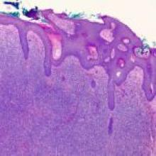
A 68-year-old woman presented to the emergency department after a family member checked on the patient and found her in unlivable home conditions. Once she arrived at the emergency department, the patient had no acute complaints, but had a large, masslike open wound that covered the upper half of the patient's face/forehead with noted infestation of maggots on the infected skin.

View Case
  • Read more about 2021apr
  • Comments
Advertisement
Subscribe to RSS - Diffuse Large B-Cell Lymphoma

Indexed Content

  • Current Issue
  • Accepted Manuscripts
  • Article Preview
  • Past Issues
  • Editorials
  • Editor's Choice
  • Fellows' Journal Club
  • Letters to the Editor
  • Video Articles

Cases

  • Case Collection
  • Archive - Case of the Week
  • Archive - Case of the Month
  • Archive - Classic Case

More from AJNR

  • Trainee Corner
  • Imaging Protocols
  • MRI Safety Corner
  • Book Reviews

Multimedia

  • AJNR Podcasts
  • AJNR Scantastics

Resources

  • Turnaround Time
  • Submit a Manuscript
  • Submit a Video Article
  • Submit an eLetter to the Editor/Response
  • Manuscript Submission Guidelines
  • Statistical Tips
  • Fast Publishing of Accepted Manuscripts
  • Graphical Abstract Preparation
  • Imaging Protocol Submission
  • Evidence-Based Medicine Level Guide
  • Publishing Checklists
  • Author Policies
  • Become a Reviewer/Academy of Reviewers
  • News and Updates

About Us

  • About AJNR
  • Editorial Board
  • Editorial Board Alumni
  • Alerts
  • Permissions
  • Not an AJNR Subscriber? Join Now
  • Advertise with Us
  • Librarian Resources
  • Feedback
  • Terms and Conditions
  • AJNR Editorial Board Alumni

American Society of Neuroradiology

  • Not an ASNR Member? Join Now

© 2025 by the American Society of Neuroradiology All rights, including for text and data mining, AI training, and similar technologies, are reserved.
Print ISSN: 0195-6108 Online ISSN: 1936-959X

Powered by HighWire