Advertisement

Main menu

  • Home
  • Content
    • Current Issue
    • Accepted Manuscripts
    • Article Preview
    • Past Issue Archive
    • Video Articles
    • AJNR Case Collection
    • Case of the Week Archive
    • Case of the Month Archive
    • Classic Case Archive
  • Special Collections
    • AJNR Awards
    • Low-Field MRI
    • Alzheimer Disease
    • ASNR Foundation Special Collection
    • Photon-Counting CT
    • View All
  • Multimedia
    • AJNR Podcasts
    • AJNR SCANtastic
    • Trainee Corner
    • MRI Safety Corner
    • Imaging Protocols
  • For Authors
    • Submit a Manuscript
    • Submit a Video Article
    • Submit an eLetter to the Editor/Response
    • Manuscript Submission Guidelines
    • Statistical Tips
    • Fast Publishing of Accepted Manuscripts
    • Graphical Abstract Preparation
    • Imaging Protocol Submission
    • Author Policies
  • About Us
    • About AJNR
    • Editorial Board
    • Editorial Board Alumni
  • More
    • Become a Reviewer/Academy of Reviewers
    • Subscribers
    • Permissions
    • Alerts
    • Feedback
    • Advertisers
    • ASNR Home

User menu

  • Alerts
  • Log in

Search

  • Advanced search
American Journal of Neuroradiology

American Journal of Neuroradiology

ASHNR American Society of Functional Neuroradiology ASHNR American Society of Pediatric Neuroradiology ASSR
  • Alerts
  • Log in

Advanced Search

  • Home
  • Content
    • Current Issue
    • Accepted Manuscripts
    • Article Preview
    • Past Issue Archive
    • Video Articles
    • AJNR Case Collection
    • Case of the Week Archive
    • Case of the Month Archive
    • Classic Case Archive
  • Special Collections
    • AJNR Awards
    • Low-Field MRI
    • Alzheimer Disease
    • ASNR Foundation Special Collection
    • Photon-Counting CT
    • View All
  • Multimedia
    • AJNR Podcasts
    • AJNR SCANtastic
    • Trainee Corner
    • MRI Safety Corner
    • Imaging Protocols
  • For Authors
    • Submit a Manuscript
    • Submit a Video Article
    • Submit an eLetter to the Editor/Response
    • Manuscript Submission Guidelines
    • Statistical Tips
    • Fast Publishing of Accepted Manuscripts
    • Graphical Abstract Preparation
    • Imaging Protocol Submission
    • Author Policies
  • About Us
    • About AJNR
    • Editorial Board
    • Editorial Board Alumni
  • More
    • Become a Reviewer/Academy of Reviewers
    • Subscribers
    • Permissions
    • Alerts
    • Feedback
    • Advertisers
    • ASNR Home
  • Follow AJNR on Twitter
  • Visit AJNR on Facebook
  • Follow AJNR on Instagram
  • Join AJNR on LinkedIn
  • RSS Feeds

AJNR Awards, New Junior Editors, and more. Read the latest AJNR updates

Critical Illness–Associated Microbleeds

  • Background:
    • Cerebral microbleeds are a well-described feature of chronic hypertension, amyloid angiopathy, diffuse axonal injury, and fat embolism. In each of these disorders, microbleeds manifest in select regions of the brain.
    • A unique distribution of microbleeds has recently been described in clinical scenarios associated with critical illness.
    • In our case, the pattern and distribution of microbleeds and associated edema were similar to those described in cases of high-altitude cerebral edema, suggesting a potential pathophysiologic mechanism could be hypoxemia leading to cytotoxic and vasogenic edema and capillary failure and leakage.
    • However, it needs to be accepted that various mechanisms contribute to a common pathway of endothelial dysfunction and blood-brain barrier breakdown. Defining a specific pathophysiologic mechanism in each case is challenging.
    • Recent case series have also suggested disseminated intravascular coagulation (DIC) as a potential explanation.
  • Clinical Presentation:
    • Typically, patients are extremely unwell and in intensive care.
    • Features of DIC, thrombocytopenia, or other clinical conditions associated with DIC, such as sepsis, may coexist.
    • A similar pattern of microbleeds has been described in patients following extracorporeal membrane oxygenation, high-altitude exposure, acute respiratory distress syndrome, and, more recently, patients with COVID-19 infection.
  • Key Diagnostic Features:
    • This phenomenon is characterized by numerous diffuse small foci of susceptibility, thought to represent microbleeds, showing a predilection for the white matter tracts, with sparing of the gray matter. The typical distribution is therefore the subcortical and juxtacortical white matter and corpus callosum.
    • Involvement of the internal capsules, external capsules, middle cerebellar peduncles, and brainstem has also been described and is demonstrated in our case.
    • In our case, foci of hemorrhage were visible on CT and most areas of microbleeds demonstrated on MRI were associated with T2 and FLAIR hyperintensity, consistent with edema. These coexisting findings have not been described in previous reports. Callosal T2 hyperintensity in this particular case may reflect edema related to hemorrhage or severe hypoxic-ischemic injury.
  • Differential Diagnoses:
    • Amyloid angiopathy: The distribution of microbleeds is typically at the gray-white matter junction, with sparing of the basal ganglia and pons. Involvement of the corpus callosum has not been described. Additional features include lobar hemorrhage and convexity subarachnoid hemorrhage.
    • Chronic hypertension (hypertensive microangiopathy): The typical distribution of microbleeds is within the basal ganglia, pons, and cerebellar hemispheres. Again, the corpus callosum is not usually involved.
    • Diffuse axonal injury: Could have a very similar distribution, but a history of major trauma would be expected
    • Cerebral fat embolism: Microbleeds are diffuse and can involve the white matter, corpus callosum, brainstem, and cerebellar hemispheres. Usually, there is involvement of both the deep and subcortical white matter. Additional features to be expected are a diffuse “starfield” pattern of punctate restricted diffusion or confluent symmetric periventricular and subcortical white matter restricted diffusion.
  • Treatment:
    • ​Supportive care
September 30, 2021

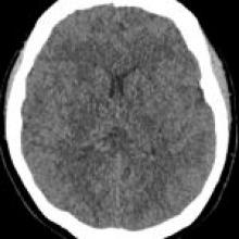
A 63-year-old woman was found unresponsive and in respiratory arrest following attempted suicide by self-asphyxiation. She was intubated and admitted directly to the ICU. MRI was performed 10 days into ICU admission due to abnormal cognition during weaning off sedation. Previous intracranial MRI, performed during an unrelated presentation, had been normal.

View Case
  • Read more about 09302021
  • Comments
Advertisement
Subscribe to RSS - Critical Illness–Associated Microbleeds

Indexed Content

  • Current Issue
  • Accepted Manuscripts
  • Article Preview
  • Past Issues
  • Editorials
  • Editor's Choice
  • Fellows' Journal Club
  • Letters to the Editor
  • Video Articles

Cases

  • Case Collection
  • Archive - Case of the Week
  • Archive - Case of the Month
  • Archive - Classic Case

More from AJNR

  • Trainee Corner
  • Imaging Protocols
  • MRI Safety Corner
  • Book Reviews

Multimedia

  • AJNR Podcasts
  • AJNR Scantastics

Resources

  • Turnaround Time
  • Submit a Manuscript
  • Submit a Video Article
  • Submit an eLetter to the Editor/Response
  • Manuscript Submission Guidelines
  • Statistical Tips
  • Fast Publishing of Accepted Manuscripts
  • Graphical Abstract Preparation
  • Imaging Protocol Submission
  • Evidence-Based Medicine Level Guide
  • Publishing Checklists
  • Author Policies
  • Become a Reviewer/Academy of Reviewers
  • News and Updates

About Us

  • About AJNR
  • Editorial Board
  • Editorial Board Alumni
  • Alerts
  • Permissions
  • Not an AJNR Subscriber? Join Now
  • Advertise with Us
  • Librarian Resources
  • Feedback
  • Terms and Conditions
  • AJNR Editorial Board Alumni

American Society of Neuroradiology

  • Not an ASNR Member? Join Now

© 2025 by the American Society of Neuroradiology All rights, including for text and data mining, AI training, and similar technologies, are reserved.
Print ISSN: 0195-6108 Online ISSN: 1936-959X

Powered by HighWire