Advertisement

Main menu

  • Home
  • Content
    • Current Issue
    • Accepted Manuscripts
    • Article Preview
    • Past Issue Archive
    • Video Articles
    • AJNR Case Collection
    • Case of the Week Archive
    • Case of the Month Archive
    • Classic Case Archive
  • Special Collections
    • AJNR Awards
    • Low-Field MRI
    • Alzheimer Disease
    • ASNR Foundation Special Collection
    • Photon-Counting CT
    • View All
  • Multimedia
    • AJNR Podcasts
    • AJNR SCANtastic
    • Trainee Corner
    • MRI Safety Corner
    • Imaging Protocols
  • For Authors
    • Submit a Manuscript
    • Submit a Video Article
    • Submit an eLetter to the Editor/Response
    • Manuscript Submission Guidelines
    • Statistical Tips
    • Fast Publishing of Accepted Manuscripts
    • Graphical Abstract Preparation
    • Imaging Protocol Submission
    • Author Policies
  • About Us
    • About AJNR
    • Editorial Board
    • Editorial Board Alumni
  • More
    • Become a Reviewer/Academy of Reviewers
    • Subscribers
    • Permissions
    • Alerts
    • Feedback
    • Advertisers
    • ASNR Home

User menu

  • Alerts
  • Log in

Search

  • Advanced search
American Journal of Neuroradiology

American Journal of Neuroradiology

ASHNR American Society of Functional Neuroradiology ASHNR American Society of Pediatric Neuroradiology ASSR
  • Alerts
  • Log in

Advanced Search

  • Home
  • Content
    • Current Issue
    • Accepted Manuscripts
    • Article Preview
    • Past Issue Archive
    • Video Articles
    • AJNR Case Collection
    • Case of the Week Archive
    • Case of the Month Archive
    • Classic Case Archive
  • Special Collections
    • AJNR Awards
    • Low-Field MRI
    • Alzheimer Disease
    • ASNR Foundation Special Collection
    • Photon-Counting CT
    • View All
  • Multimedia
    • AJNR Podcasts
    • AJNR SCANtastic
    • Trainee Corner
    • MRI Safety Corner
    • Imaging Protocols
  • For Authors
    • Submit a Manuscript
    • Submit a Video Article
    • Submit an eLetter to the Editor/Response
    • Manuscript Submission Guidelines
    • Statistical Tips
    • Fast Publishing of Accepted Manuscripts
    • Graphical Abstract Preparation
    • Imaging Protocol Submission
    • Author Policies
  • About Us
    • About AJNR
    • Editorial Board
    • Editorial Board Alumni
  • More
    • Become a Reviewer/Academy of Reviewers
    • Subscribers
    • Permissions
    • Alerts
    • Feedback
    • Advertisers
    • ASNR Home
  • Follow AJNR on Twitter
  • Visit AJNR on Facebook
  • Follow AJNR on Instagram
  • Join AJNR on LinkedIn
  • RSS Feeds

AJNR Awards, New Junior Editors, and more. Read the latest AJNR updates

Cerebral Amyloid Angiopathy–Related Inflammation (CAARI)

  • Background:
    • CAARI, also called amyloid-β-related angiitis, is a rare form of cerebral amyloid angiopathy with a predominantly vascular inflammation or angiitis.
  • Clinical Presentation:
    • Patients typically present with seizures, headache, and strokelike episodes, along with an acute or subacute decline in cognitive status.
  • Key Diagnostic Features:
    • The striking radiologic features include vasogenic edema involving the subcortical white matter, accompanied by mass effect, typically in frontal and temporal lobes, but presenting as a multifocal involvement of both hemispheres in 30% of patients. Cortical involvement can occur and accounts for seizures in these patients. These changes in association with multiple microhemorrhages diffusely distributed throughout the brain (best seen on SWI) make the diagnosis.
    • Slight leptomeningeal enhancement can also be present.
    • A reliable diagnosis of CAARI can be reached from clinical and MRI information alone, with good sensitivity and excellent specificity, exempting brain biopsy.
    • According to new criteria, probable CAARI will be diagnosed in the setting of the following:
      • Age ≥ 40 years
      • Acute or subacute onset of symptoms
      • Presence of ≥ 1 of the following clinical features: headache, decrease in consciousness, behavioral change, or focal neurologic signs and seizures; the presentation is not directly attributable to an acute ICH
      • MRI shows unifocal or multifocal WMH lesions (corticosubcortical or deep) that are asymmetric and extend to the immediately subcortical white matter; the asymmetry is not due to past ICH
      • Presence of ≥ 1 of the following corticosubcortical hemorrhagic lesions: cerebral macrobleed, cerebral microbleed, or cortical superficial siderosis
      • Absence of neoplastic, infectious, or other cause
  • Differential Diagnoses
    • The main differential diagnosis includes primary CNS vasculitis, PRES, and PML. When there is one single lesion, brain tumor or tumefactive inflammatory lesion may also be considered in the differential.
  • Treatment:
    • These patients usually respond well to a pulse of corticosteroids.
    • Some authors have stated the role of immune or cytostatic agents.
July 2020

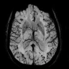
A 77-year-old man with sudden onset of mild sensitive aphasia and no other neurologic symptoms. His history included 3 previous, similar but shorter episodes and slowly progressive mental status deterioration in the last 2 months. Besides the aphasia, the neurologic examination was unremarkable. Blood tests were normal and CSF showed high protein without glucose consumption.

View Case
  • Read more about 2020jul
  • Comments
Advertisement
Subscribe to RSS - Cerebral Amyloid Angiopathy–Related Inflammation (CAARI)

Indexed Content

  • Current Issue
  • Accepted Manuscripts
  • Article Preview
  • Past Issues
  • Editorials
  • Editor's Choice
  • Fellows' Journal Club
  • Letters to the Editor
  • Video Articles

Cases

  • Case Collection
  • Archive - Case of the Week
  • Archive - Case of the Month
  • Archive - Classic Case

More from AJNR

  • Trainee Corner
  • Imaging Protocols
  • MRI Safety Corner
  • Book Reviews

Multimedia

  • AJNR Podcasts
  • AJNR Scantastics

Resources

  • Turnaround Time
  • Submit a Manuscript
  • Submit a Video Article
  • Submit an eLetter to the Editor/Response
  • Manuscript Submission Guidelines
  • Statistical Tips
  • Fast Publishing of Accepted Manuscripts
  • Graphical Abstract Preparation
  • Imaging Protocol Submission
  • Evidence-Based Medicine Level Guide
  • Publishing Checklists
  • Author Policies
  • Become a Reviewer/Academy of Reviewers
  • News and Updates

About Us

  • About AJNR
  • Editorial Board
  • Editorial Board Alumni
  • Alerts
  • Permissions
  • Not an AJNR Subscriber? Join Now
  • Advertise with Us
  • Librarian Resources
  • Feedback
  • Terms and Conditions
  • AJNR Editorial Board Alumni

American Society of Neuroradiology

  • Not an ASNR Member? Join Now

© 2025 by the American Society of Neuroradiology All rights, including for text and data mining, AI training, and similar technologies, are reserved.
Print ISSN: 0195-6108 Online ISSN: 1936-959X

Powered by HighWire