Advertisement

Main menu

  • Home
  • Content
    • Current Issue
    • Accepted Manuscripts
    • Article Preview
    • Past Issue Archive
    • Video Articles
    • AJNR Case Collection
    • Case of the Week Archive
    • Case of the Month Archive
    • Classic Case Archive
  • Special Collections
    • AJNR Awards
    • Low-Field MRI
    • Alzheimer Disease
    • ASNR Foundation Special Collection
    • Photon-Counting CT
    • View All
  • Multimedia
    • AJNR Podcasts
    • AJNR SCANtastic
    • Trainee Corner
    • MRI Safety Corner
    • Imaging Protocols
  • For Authors
    • Submit a Manuscript
    • Submit a Video Article
    • Submit an eLetter to the Editor/Response
    • Manuscript Submission Guidelines
    • Statistical Tips
    • Fast Publishing of Accepted Manuscripts
    • Graphical Abstract Preparation
    • Imaging Protocol Submission
    • Author Policies
  • About Us
    • About AJNR
    • Editorial Board
    • Editorial Board Alumni
  • More
    • Become a Reviewer/Academy of Reviewers
    • Subscribers
    • Permissions
    • Alerts
    • Feedback
    • Advertisers
    • ASNR Home

User menu

  • Alerts
  • Log in

Search

  • Advanced search
American Journal of Neuroradiology

American Journal of Neuroradiology

ASHNR American Society of Functional Neuroradiology ASHNR American Society of Pediatric Neuroradiology ASSR
  • Alerts
  • Log in

Advanced Search

  • Home
  • Content
    • Current Issue
    • Accepted Manuscripts
    • Article Preview
    • Past Issue Archive
    • Video Articles
    • AJNR Case Collection
    • Case of the Week Archive
    • Case of the Month Archive
    • Classic Case Archive
  • Special Collections
    • AJNR Awards
    • Low-Field MRI
    • Alzheimer Disease
    • ASNR Foundation Special Collection
    • Photon-Counting CT
    • View All
  • Multimedia
    • AJNR Podcasts
    • AJNR SCANtastic
    • Trainee Corner
    • MRI Safety Corner
    • Imaging Protocols
  • For Authors
    • Submit a Manuscript
    • Submit a Video Article
    • Submit an eLetter to the Editor/Response
    • Manuscript Submission Guidelines
    • Statistical Tips
    • Fast Publishing of Accepted Manuscripts
    • Graphical Abstract Preparation
    • Imaging Protocol Submission
    • Author Policies
  • About Us
    • About AJNR
    • Editorial Board
    • Editorial Board Alumni
  • More
    • Become a Reviewer/Academy of Reviewers
    • Subscribers
    • Permissions
    • Alerts
    • Feedback
    • Advertisers
    • ASNR Home
  • Follow AJNR on Twitter
  • Visit AJNR on Facebook
  • Follow AJNR on Instagram
  • Join AJNR on LinkedIn
  • RSS Feeds

AJNR Awards, New Junior Editors, and more. Read the latest AJNR updates

Research ArticleNEUROVASCULAR/STROKE IMAGING

Identification of Multiclass Pediatric Low-Grade Neuroepithelial Tumor Molecular Subtype with ADC MR Imaging and Machine Learning

Matheus D. Soldatelli, Khashayar Namdar, Uri Tabori, Cynthia Hawkins, Kristen Yeom, Farzad Khalvati, Birgit B. Ertl-Wagner and Matthias W. Wagner
American Journal of Neuroradiology April 2024, DOI: https://doi.org/10.3174/ajnr.A8199
Matheus D. Soldatelli
aFrom the Department Diagnostic Imaging (M.D.S., B.B.E.-W., M.W.W.), Division of Neuroradiology, The Hospital for Sick Children, Toronto, Ontario, Canada
bDepartment of Medical Imaging (M.D.S., K.N., F.K., B.B.E.-W., M.W.W.), University of Toronto, Toronto, Ontario, Canada
cInstitute of Medical Science (M.D.S., K.N., U.T., F.K., B.B.E.-W.), University of Toronto, Toronto, Ontario, Canada
  • Find this author on Google Scholar
  • Find this author on PubMed
  • Search for this author on this site
  • ORCID record for Matheus D. Soldatelli
Khashayar Namdar
bDepartment of Medical Imaging (M.D.S., K.N., F.K., B.B.E.-W., M.W.W.), University of Toronto, Toronto, Ontario, Canada
cInstitute of Medical Science (M.D.S., K.N., U.T., F.K., B.B.E.-W.), University of Toronto, Toronto, Ontario, Canada
dVector Institute (K.N., F.K.), Toronto, Ontario, Canada
  • Find this author on Google Scholar
  • Find this author on PubMed
  • Search for this author on this site
Uri Tabori
cInstitute of Medical Science (M.D.S., K.N., U.T., F.K., B.B.E.-W.), University of Toronto, Toronto, Ontario, Canada
eThe Arthur and Sonia Labatt Brain Tumour Research Centre (U.T., C.H.), The Hospital for Sick Children, Toronto, Ontario, Canada
fProgram in Genetics and Genome Biology (U.T.) The Hospital for Sick Children, Toronto, Ontario, Canada
  • Find this author on Google Scholar
  • Find this author on PubMed
  • Search for this author on this site
  • ORCID record for Uri Tabori
Cynthia Hawkins
eThe Arthur and Sonia Labatt Brain Tumour Research Centre (U.T., C.H.), The Hospital for Sick Children, Toronto, Ontario, Canada
gDepartment of Laboratory Medicine and Pathobiology (C.H.), University of Toronto, Toronto, Ontario, Canada
hDivision of Pathology (C.H.), The Hospital for Sick Children, Toronto, Ontario, Canada
  • Find this author on Google Scholar
  • Find this author on PubMed
  • Search for this author on this site
  • ORCID record for Cynthia Hawkins
Kristen Yeom
iDepartment of Radiology (K.Y.), Lucile Packard Children's Hospital, Stanford University School of Medicine, Stanford, California
  • Find this author on Google Scholar
  • Find this author on PubMed
  • Search for this author on this site
  • ORCID record for Kristen Yeom
Farzad Khalvati
bDepartment of Medical Imaging (M.D.S., K.N., F.K., B.B.E.-W., M.W.W.), University of Toronto, Toronto, Ontario, Canada
cInstitute of Medical Science (M.D.S., K.N., U.T., F.K., B.B.E.-W.), University of Toronto, Toronto, Ontario, Canada
dVector Institute (K.N., F.K.), Toronto, Ontario, Canada
jDepartment of Computer Science (F.K.), University of Toronto, Toronto, Ontario, Canada
  • Find this author on Google Scholar
  • Find this author on PubMed
  • Search for this author on this site
  • ORCID record for Farzad Khalvati
Birgit B. Ertl-Wagner
aFrom the Department Diagnostic Imaging (M.D.S., B.B.E.-W., M.W.W.), Division of Neuroradiology, The Hospital for Sick Children, Toronto, Ontario, Canada
bDepartment of Medical Imaging (M.D.S., K.N., F.K., B.B.E.-W., M.W.W.), University of Toronto, Toronto, Ontario, Canada
cInstitute of Medical Science (M.D.S., K.N., U.T., F.K., B.B.E.-W.), University of Toronto, Toronto, Ontario, Canada
  • Find this author on Google Scholar
  • Find this author on PubMed
  • Search for this author on this site
  • ORCID record for Birgit B. Ertl-Wagner
Matthias W. Wagner
aFrom the Department Diagnostic Imaging (M.D.S., B.B.E.-W., M.W.W.), Division of Neuroradiology, The Hospital for Sick Children, Toronto, Ontario, Canada
bDepartment of Medical Imaging (M.D.S., K.N., F.K., B.B.E.-W., M.W.W.), University of Toronto, Toronto, Ontario, Canada
kDepartment of Diagnostic and Interventional Neuroradiology (M.W.W.), University Hospital Augsburg, Augsburg, Germany
  • Find this author on Google Scholar
  • Find this author on PubMed
  • Search for this author on this site
  • ORCID record for Matthias W. Wagner
  • Article
  • Figures & Data
  • Info & Metrics
  • Responses
  • References
  • PDF
Loading

Article Figures & Data

Figures

  • Tables
  • FIG 1.
    • Download figure
    • Open in new tab
    • Download powerpoint
    FIG 1.

    Axial FLAIR (A and D), ADC maps (B and E), and manual tumor segmentation using semiautomatic tools in 3D Slicer (C and F). Upper row: a 15-year-old adolescent boy with a supratentorial BRAF-mutated low-grade glioma. Lower row: a 3-year-old boy with a cerebellar BRAF-fused low-grade glioma.

  • FIG 2.
    • Download figure
    • Open in new tab
    • Download powerpoint
    FIG 2.

    Flow chart of the study.
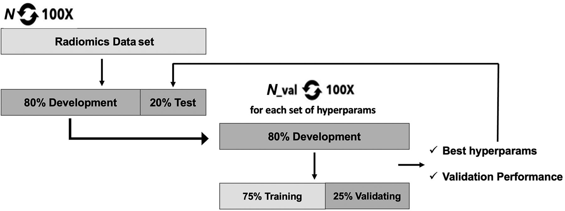
  • FIG 3.
    • Download figure
    • Open in new tab
    • Download powerpoint
    FIG 3.

    The repetitive classification approach.
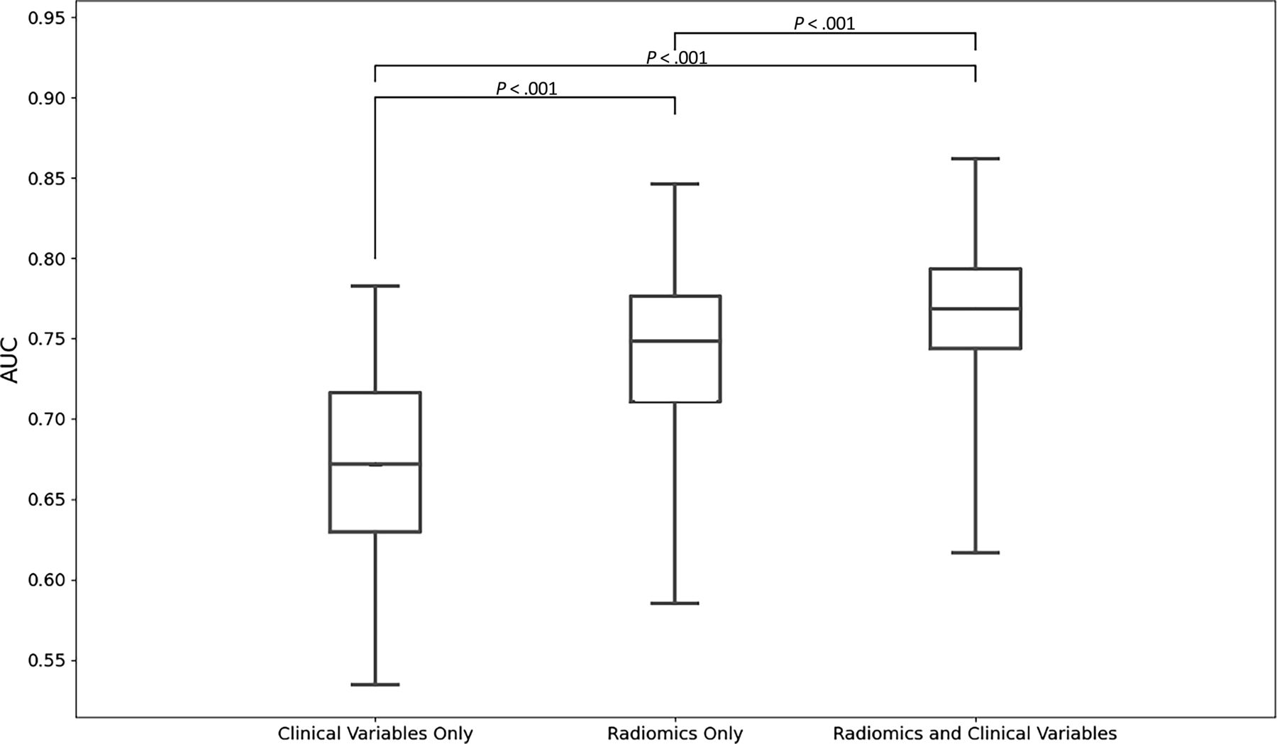
  • FIG 4.
    • Download figure
    • Open in new tab
    • Download powerpoint
    FIG 4.

    Boxplots of test AUCs. Note that P values are calculated on the basis of Student t tests.

Tables

  • Figures
    • View popup
    Table 1:

    Patient demographics

    Institutional CohortTotal
    TorontoStanford
    No. of patients22923252
    Age (mean) (yr)8.186.328.01
    Male (No.) (%)124 (54.1%)11 (47.8%)135 (53.6%)
    Histologic diagnosis (No.)
     PA11216128
     GG24529
     LGA36036
     PMA8210
     PXA404
     DNET10010
     DA17017
     ODG303
     GNT505
     ACG505
     NC101
     DIG101
     PLNTY101
    Mixed tumor components202
    Molecular subgroup (No.) (%)
     KIAA1549-BRAF fusion114 (49.7%)18 (78.3%)132 (52.2%)
     BRAF V600E mutation36 (15.7%)5 (21.7%)41 (16.6%)
     Non-BRAF79 (34.4%)079 (31.2%)
     Supratentorial (No.) (%)125 (54.5%)5 (21.7%)130 (51.8%)
     Infratentorial (No.) (%)104 (45.4%)18 (78.3%)122 (48.2%)
    • Note:—ACG indicates angiocentric glioma; DA, diffuse astrocytoma; DIG, desmoplastic infantile ganglioglioma; DNET, dysembryoplastic neuroepithelial tumor; GNT, glioneural tumor; LGA, low-grade astrocytoma; NC, neurocytoma; PMA, pilomyxoid astrocytoma; PXA, pleomorphic xanthoastrocytoma; PLNTY, polymorphous low-grade neuroepithelial tumor of the young; ODG, oligodendroglioma.

    • View popup
    Table 2:

    Top predictive radiomics and clinical features

    TypeSourceFeature CategoryFeature
    ClinicalNANATumor location
    Radiomics3D wavelet transformGray-level difference matrixLarge dependence high gray-level emphasis
    Radiomics3D wavelet transformFirst orderMean
    Radiomics3D wavelet transformFirst orderKurtosis
    RadiomicsOriginalGray-level difference matrixDependence variance
    RadiomicsOriginalGray-level difference matrixLarge dependence high gray-level emphasis
    RadiomicsLogarithmGray-level difference matrixLarge dependence high gray-level emphasis
    RadiomicsLocal binary pattern 3DGray-level difference matrixLarge dependence emphasis
    RadiomicsExponentialFirst order90th Percentile
    Radiomics3D wavelet transformFirst orderKurtosis
    • Note:—NA indicates not applicable.

    • View popup
    Table 3:

    Per-class and overall OvR test of AUC performance of the models

    KIAA1549-BRAF Fusion versus the RestBRAF V600E Mutation versus the RestNon-BRAF versus the RestAverage
    Radiomics features onlyAUC = 0.80 95% CI, 0.79–0.81AUC = 0.74 95% CI, 0.73–0.75AUC = 0.67 95% CI, 0.66–0.68AUC = 0.74 95% CI, 0.73–0.75
    Clinical features onlyAUC = 0.75 95% CI, 0.74–0.7AUC = 0.62 95% CI, 0.61–0.63AUC = 0.63 95% CI, 0.62–0.6AUC = 0.67 95% CI, 0.66–0.68
    Combined radiomics and clinical featuresAUC = 0.81 95% CI, 0.81–0.82AUC = 0.75 95% CI, 0.73–0.75AUC = 0.74 95% CI, 0.672–0.74AUC = 0.76 95% CI, 0.75–0.7)
PreviousNext
Back to top
Advertisement
Print
Download PDF
Email Article

Thank you for your interest in spreading the word on American Journal of Neuroradiology.

NOTE: We only request your email address so that the person you are recommending the page to knows that you wanted them to see it, and that it is not junk mail. We do not capture any email address.

Enter multiple addresses on separate lines or separate them with commas.
Identification of Multiclass Pediatric Low-Grade Neuroepithelial Tumor Molecular Subtype with ADC MR Imaging and Machine Learning
(Your Name) has sent you a message from American Journal of Neuroradiology
(Your Name) thought you would like to see the American Journal of Neuroradiology web site.
CAPTCHA
This question is for testing whether or not you are a human visitor and to prevent automated spam submissions.
Cite this article
Matheus D. Soldatelli, Khashayar Namdar, Uri Tabori, Cynthia Hawkins, Kristen Yeom, Farzad Khalvati, Birgit B. Ertl-Wagner, Matthias W. Wagner
Identification of Multiclass Pediatric Low-Grade Neuroepithelial Tumor Molecular Subtype with ADC MR Imaging and Machine Learning
American Journal of Neuroradiology Apr 2024, DOI: 10.3174/ajnr.A8199

Citation Manager Formats

  • BibTeX
  • Bookends
  • EasyBib
  • EndNote (tagged)
  • EndNote 8 (xml)
  • Medlars
  • Mendeley
  • Papers
  • RefWorks Tagged
  • Ref Manager
  • RIS
  • Zotero
0 Responses
Respond to this article
Share
Bookmark this article
Identification of Multiclass Pediatric Low-Grade Neuroepithelial Tumor Molecular Subtype with ADC MR Imaging and Machine Learning
Matheus D. Soldatelli, Khashayar Namdar, Uri Tabori, Cynthia Hawkins, Kristen Yeom, Farzad Khalvati, Birgit B. Ertl-Wagner, Matthias W. Wagner
American Journal of Neuroradiology Apr 2024, DOI: 10.3174/ajnr.A8199
del.icio.us logo Twitter logo Facebook logo Mendeley logo
  • Tweet Widget
  • Facebook Like
  • Google Plus One
Purchase

Jump to section

  • Article
    • Abstract
    • ABBREVIATIONS:
    • MATERIALS AND METHODS
    • RESULTS
    • DISCUSSION
    • CONCLUSIONS
    • Footnotes
    • References
  • Figures & Data
  • Info & Metrics
  • Responses
  • References
  • PDF

Related Articles

  • No related articles found.
  • PubMed
  • Google Scholar

Cited By...

  • Segmentation-Free Pretherapeutic Assessment of BRAF-Status in Pediatric Low-Grade Gliomas
  • Crossref
  • Google Scholar

This article has not yet been cited by articles in journals that are participating in Crossref Cited-by Linking.

More in this TOC Section

  • The insula and malignant cerebral edema
  • ICH Shape & Density Predict Rebleeding after MICE
  • Circle of Willis Variants and Stroke Outcomes
Show more Neurovascular/Stroke Imaging

Similar Articles

Advertisement

Indexed Content

  • Current Issue
  • Accepted Manuscripts
  • Article Preview
  • Past Issues
  • Editorials
  • Editor's Choice
  • Fellows' Journal Club
  • Letters to the Editor
  • Video Articles

Cases

  • Case Collection
  • Archive - Case of the Week
  • Archive - Case of the Month
  • Archive - Classic Case

More from AJNR

  • Trainee Corner
  • Imaging Protocols
  • MRI Safety Corner
  • Book Reviews

Multimedia

  • AJNR Podcasts
  • AJNR Scantastics

Resources

  • Turnaround Time
  • Submit a Manuscript
  • Submit a Video Article
  • Submit an eLetter to the Editor/Response
  • Manuscript Submission Guidelines
  • Statistical Tips
  • Fast Publishing of Accepted Manuscripts
  • Graphical Abstract Preparation
  • Imaging Protocol Submission
  • Evidence-Based Medicine Level Guide
  • Publishing Checklists
  • Author Policies
  • Become a Reviewer/Academy of Reviewers
  • News and Updates

About Us

  • About AJNR
  • Editorial Board
  • Editorial Board Alumni
  • Alerts
  • Permissions
  • Not an AJNR Subscriber? Join Now
  • Advertise with Us
  • Librarian Resources
  • Feedback
  • Terms and Conditions
  • AJNR Editorial Board Alumni

American Society of Neuroradiology

  • Not an ASNR Member? Join Now

© 2025 by the American Society of Neuroradiology All rights, including for text and data mining, AI training, and similar technologies, are reserved.
Print ISSN: 0195-6108 Online ISSN: 1936-959X

Powered by HighWire