
Article Figures & Data
Figures
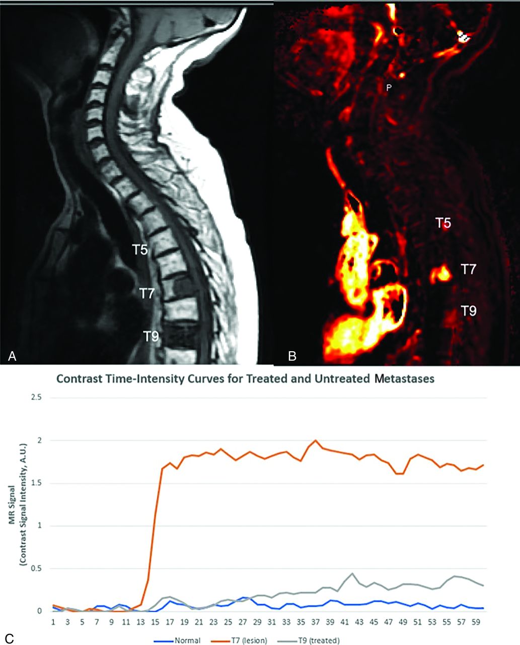
- FIG 1.
Graphic of normal marrow, viable tumor, and treated metastatic breast carcinoma. A, Sagittal T1 precontrast image demonstrates normal marrow signal at T5, posterior T7 metastasis, and diffuse metastatic involvement of T9. Based on the sagittal T1-precontrast (shown) and the T2 and postcontrast T1 images (not shown), it is not possible to distinguish between viable tumor and successfully treated metastasis. B, Perfusion imaging illustrates elevated plasma volume correlating to T7 metastasis (bright orange color) consistent with viable tumor. There is no hyperperfusion associated at T5 consistent with normal marrow as well as with T9 consistent with treated metastasis. C, TICs demonstrate rapid wash-in and plateau (type C curve) for T7 lesion consistent with viable tumor, while normal T5 and treated tumor at T9 demonstrate relatively flat (type A) TIC.
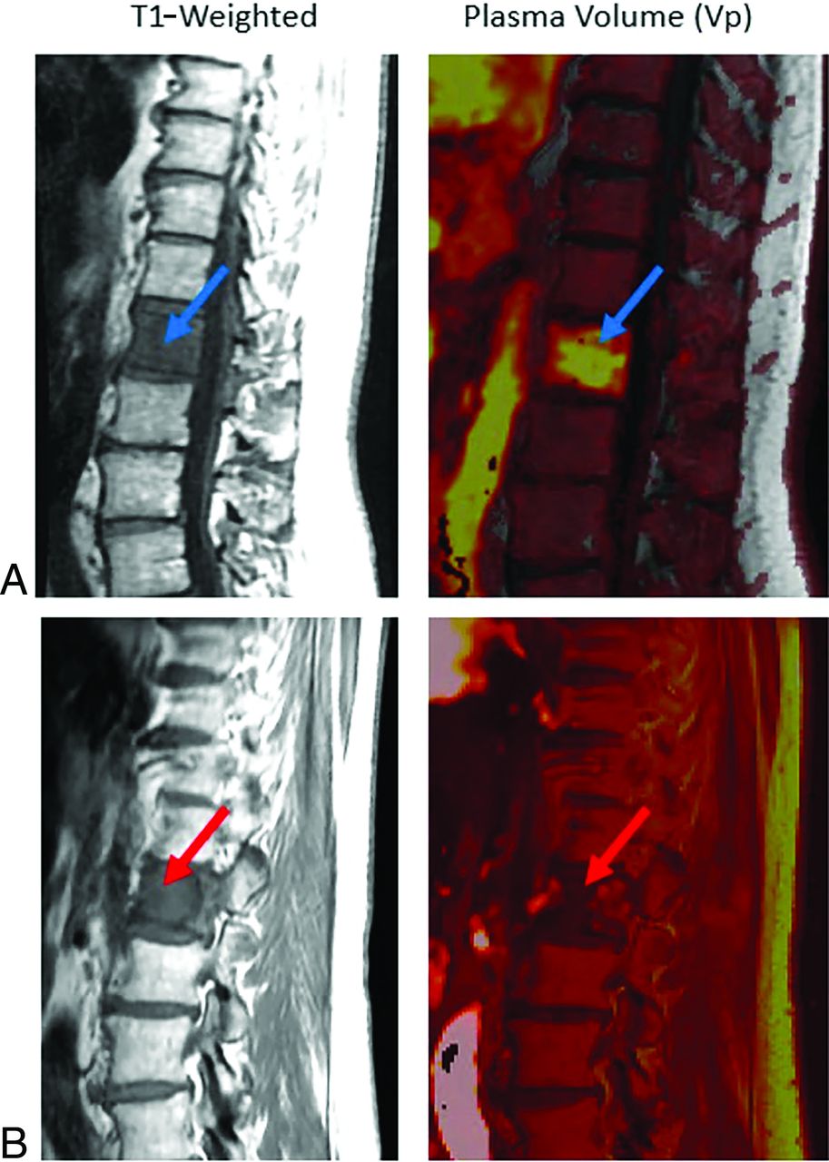
- FIG 2.
Non-neoplastic versus pathologic compression fractures. A, Sagittal T1-weighted image demonstrates diffuse hypointense marrow replacing lesion within L1 consistent with breast carcinoma (blue arrow), with mild vertebral height loss consistent with compression fracture. There is expansion into the ventral epidural space in the setting of correlative elevated plasma volume on perfusion imaging (bright yellow on color map; green arrow), which suggests viable tumor. Constellation of findings is consistent with pathologic fracture. B, Sagittal T1 demonstrates hypointense lesion at superior T9 (blue arrow) without associated elevated plasma volume (dark signal on color map; green arrow) in a patient with lung carcinoma. Constellation of findings is consistent with non-neoplastic compression fracture.
- FIG 3.
Metastatic disease versus atypical hemangioma. Sagittal images demonstrate diffuse STIR hyperintense L1 lesion (top left, blue arrow) and smaller STIR hyperintense L3 lesion (gray arrow). Both lesions demonstrate T1 hypointensity (middle). Perfusion imaging (top right) demonstrates elevated plasma volume associated with the L1 lesion, which is suggestive of metastasis (subsequently, biopsy proved to be metastatic gastrointestinal tumor), and reduced plasma volume for the L3 lesion, which is suggestive of non-neoplastic etiology. In conjunction with anatomic appearance, the L3 lesion is most consistent with an atypical hemangioma. Bottom, The L1 lesion demonstrates type C TIC consistent with metastasis, and L3 lesion demonstrates flat TIC curve consistent with non-neoplastic lesion.
- FIG 4.
Patient with metastatic colon adenocarcinoma. A, Pretreatment scan that demonstrates T1 hypointense L1 lesion (top left, blue arrow) with associated elevated plasma volume (bright yellow on color map; top right, blue arrow) of 5.84, consistent with metastatic disease. B, A 2-month postradiation therapy scan that shows a near similar appearance of T1 hypointense lesion (bottom left, red arrow) but substantial decrease in associated plasma volume (dark signal on color map; bottom right, red arrow) of 1.65, which is a 72% reduction. Constellation of findings reflect treated disease despite similar appearance on anatomic T1-weighted images of pre- and posttreatment scans.
- FIG 5.
Patient with metastatic lung adenocarcinoma to L4. A and B were obtained several months apart demonstrating T1 hypointense marrow replacing lesion involving L4 consistent with metastatic disease. A, First posttreatment scan after the patient received radiation 7 months prior. Top left, Metastasis at L4 (blue arrow). Top right, Lack of elevated plasma volume (dark signal on color map; blue arrow). B, Posttreatment scan 10 months subsequent to A and again with a similar T1 hypointense lesion in L4 (bottom left, red arrow). However, it is difficult to delineate simply based on anatomic imaging if the lesion remains viable or not, as both demonstrate similar T1 hypointensity. The L4 lesion now demonstrates correlative elevated plasma volume (bright signal on color map; bottom left, red arrow), which is consistent with a now-viable tumor reflecting recurrent disease.
- FIG 6.
Pre- and posttreatment assessment of chordoma. A, Pretreatment sagittal images (top row) with T1 hypointense (top left - blue arrow) and STIR hyperintense (top middle - blue arrow) marrow replacing S1–S4 lesion asymmetric to left sacral ala with associated hyperperfusion (bright signal on top right - blue arrow). B, Postradiation treatment sagittal images that show similar T1 hypointense (bottom left - red arrow) and STIR hyperintense mass (bottom middle - red arrow); however, there is significant decrease in perfusion (bottom right - red arrow) consistent with treatment response. TIC curves also demonstrate reduced TIC for treated disease.


Jump to section
Related Articles
Cited By...
- No citing articles found.