
Article Figures & Data
Figures
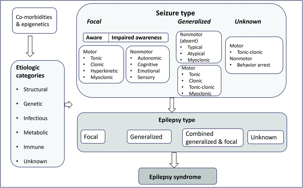
- FIG 1.
This diagram highlights the 3-tier approach to classifying disease into seizure type, epilepsy type, and, finally, the presence of any epilepsy syndrome. The major etiologic categories of epilepsy/seizures are also depicted.
- FIG 2.
Right superior frontal focal cortical dysplasia (ILAE IIb). High-resolution T2 coronal image (A) shows moderate focal cortical thickening of the right superior frontal gyrus with blurring of the gray-white matter interface (arrow). An [18F] FDG-PET scan of the brain and the fused PET/MR images (B and C) reveal focal hypometabolism corresponding to the dysplastic cortex (arrows). MEG scan (D) shows tight dipole clustering, corresponding to the site of PET and abnormal findings on MR imaging.
- FIG 3.
Ultra-high-field (7T) MR imaging scan with BOS FCD ILAE type IIa with a DEPDC5 mutation. Mild cortical thickening is noted at the base of left superior frontal sulcus with blurring of the gray-white matter interface and minimal T2 hyperintensity in the adjacent WM (arrows). The crown of the gyrus is normal. Newer sequences like 3D EDGE (D) can better define the gray-white interface, lost in the region of FCD, as seen here (D, arrow).
- FIG 4.
NeuN immunohistochemistry highlighting the neuronal cell in the normal cortex and FCDs I–III. Normal 6-layer architecture is depicted in the normal cortex (A). FCD Ia (B) shows abundant neuronal microcolumns. Low-power magnification of the cortex in FCD IIb shows complete loss of layering and many enlarged, clustered dysmorphic neurons (C, black arrows). Balloon cells are not clearly delineated on NeuN immunohistochemistry and are better seen on stains like crystallin (see Fig 5D). FCD IIId in a young adult with a remote ischemic insult reveals cortical thinning with marked loss of neurons in the middle layer 4 (D, arrow).
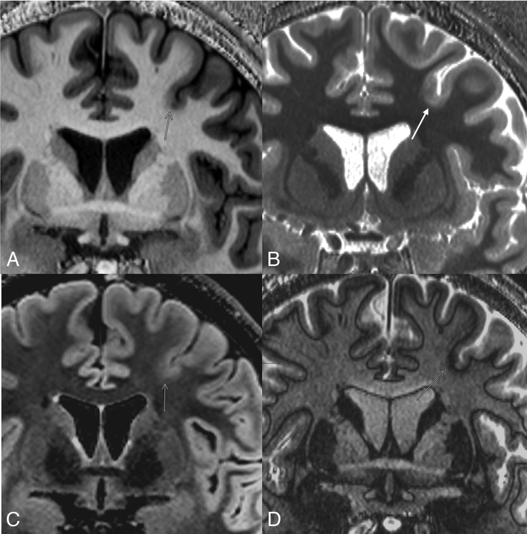
- FIG 5.
Right frontal ILAE type IIb in a 30-year-old patient with focal partial symptomatic epilepsy and epileptic syndrome. Coronal 3D EDGE (A) and T1-gradient-echo image (MP2RAGE) (B) obtained on a 7T magnet reveal cortical thickening with blurring of gray-white matter interface (arrows). Note a transmantle band reaching up to the ventricle margins, best seen on the T2 FLAIR coronal image (C, arrow). MP2RAGE depicts the thickening and blurring of the cortex at the site of the FCD with sharp gray-white demarcation at the normal cortices, showing the superiority of T1-weighted gradient sequences for anatomic delineation. Multiple balloon cells in FCD IIb on crystallin staining of the dysplastic cortex are seen as large, round cells with oval, eccentric nuclei; prominent nucleoli; and abundant cytoplasm scattered throughout the GM (D, black arrows).
- FIG 6.
Left frontal ILAE type IIIb in an 18-year-old patient in conjunction with multinodular and vacuolating neuronal tumor (MVNT). A cluster of multinodular T1-hypointense and T2 FLAIR-hyperintense nodules (A, B, and D, white arrows) is seen in the left posterior peri-Sylvian region (supra-Sylvian perirolandic and posterior insula) consistent with MVNT. Note a dysplastic adjacent cortex involving the inferior frontal gyrus (C, black arrow) with cortical thickening and blurring of the gray-white matter interface.
- FIG 7.
MOGHE in a 2-year-old boy with surgical pathology confirmation. Multiple T2 FLAIR images (A–C) reveal focal subcortical WM hyperintensity in the right middle frontal gyrus region (arrows). Coronal T1 gradient-echo image (MPRAGE) (D) shows a normal overlying cortex with subtle hypointensity (arrow) corresponding to the WM signal changes seen on T2/FLAIR. A right frontal resection was performed with excellent seizure control. Histopathology revealed subcortical WM hypercellularity, largely oligodendroglial (on Olig-2 stains), suggestive of oligodendroglial hyperplasia. NeuN highlighted a few scattered neurons in the WM; however, no dysmorphic neurons, balloon cells, or cortical dysplasia was noted.


Tables
Comparison of Blumcke 2011 classification of FCDs with the updated ILAE 2022 classification, along with the new 2022 entities
Blumcke (ILAE 2011) ILAE 2022 FCD type I, isolated focal cortical dysplasia Ia abnormal radial cortical lamination Ia abundant neuronal microcolumns (vertical) Ib abnormal tangential cortical lamination Ib abnormal tangential layering Ic abnormal radial and tangential cortical lamination Ic vertical and horizontal abnormalities FCD type II, isolated, focal, cortical dysplasia IIa dysmorphic neurons IIa dysmorphic neurons IIb dysmorphic neurons and balloon cells (Taylor type) IIb dysmorphic neurons and balloon cells (Taylor type) BOCa FCD (new entity) could be IIa or IIb on histopathology) FCD type III, cortical dyslamination and principal lesion IIIa with hippocampal sclerosis IIIa with hippocampal sclerosis IIIb adjacent to glial/glioneuronal tumor IIIb adjacent to glial/glioneuronal tumor IIIc adjacent to vascular malformation IIIc adjacent to vascular malformation IIId adjacent to early life insult like ischemia IIId adjacent to early-life insult-like ischemia Term of “not otherwise specified (NOS)” should be used if microscopic diagnosis is not based on appropriate immunohistochemical staining, eg, FCD type II (NOS) New entities mMCD increase in heterotopic neurons in the WM MOGHE indicates mild malformations of cortical development with oligodendroglial hyperplasia (>2200 Olig-2 cells/mm) No definite FCD on histopathology, ambiguous pathologic findings, not compatible to FCD I or II ↵a Common immunohistochemical staining used for FCD diagnosis includes antibodies directed against NeuN neurofilaments, vimentin, MAP2, CD34, OLIG2, glial fibrillary acid protein, or α B-crystallin.


Jump to section
Related Articles
Cited By...
- No citing articles found.