
Article Figures & Data
Figures
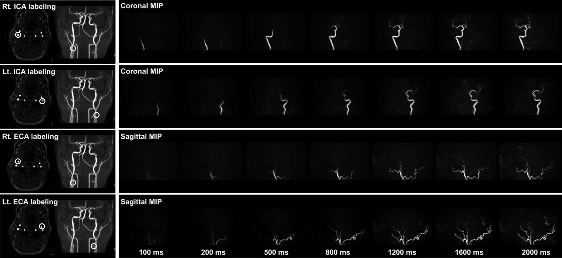
- FIG 1.
The locations of labeling spots. The labeling focus is placed in the proximal portions of the ICA and ECA within 3 cm of the bifurcation of the common carotid artery. For the labeling of each artery, 0.75 mT/m/ms is set as the gradient moment for superselective pCASL in both the right-to-left and anterior-to-posterior directions, creating a circular labeling spot with an approximately 2-cm diameter. Rt. indicates right; Lt., left.
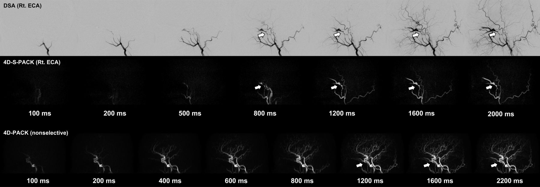
- FIG 2.
A 27-year-old woman with an intracranial DAVF at the cavernous sinus that is supplied by the right accessory meningeal artery (white arrow). The 4D-S-PACK (middle row) labeling of the right ECA clearly depicts the feeding artery (accessory meningeal artery) and draining veins (cavernous sinus and ophthalmic veins) as seen on the patient’s DSA (upper row). In contrast, it is difficult to identify the feeding artery on 4D-PACK (lower row) because of the overlap of many other unrelated vessels. Rt. indicates right.
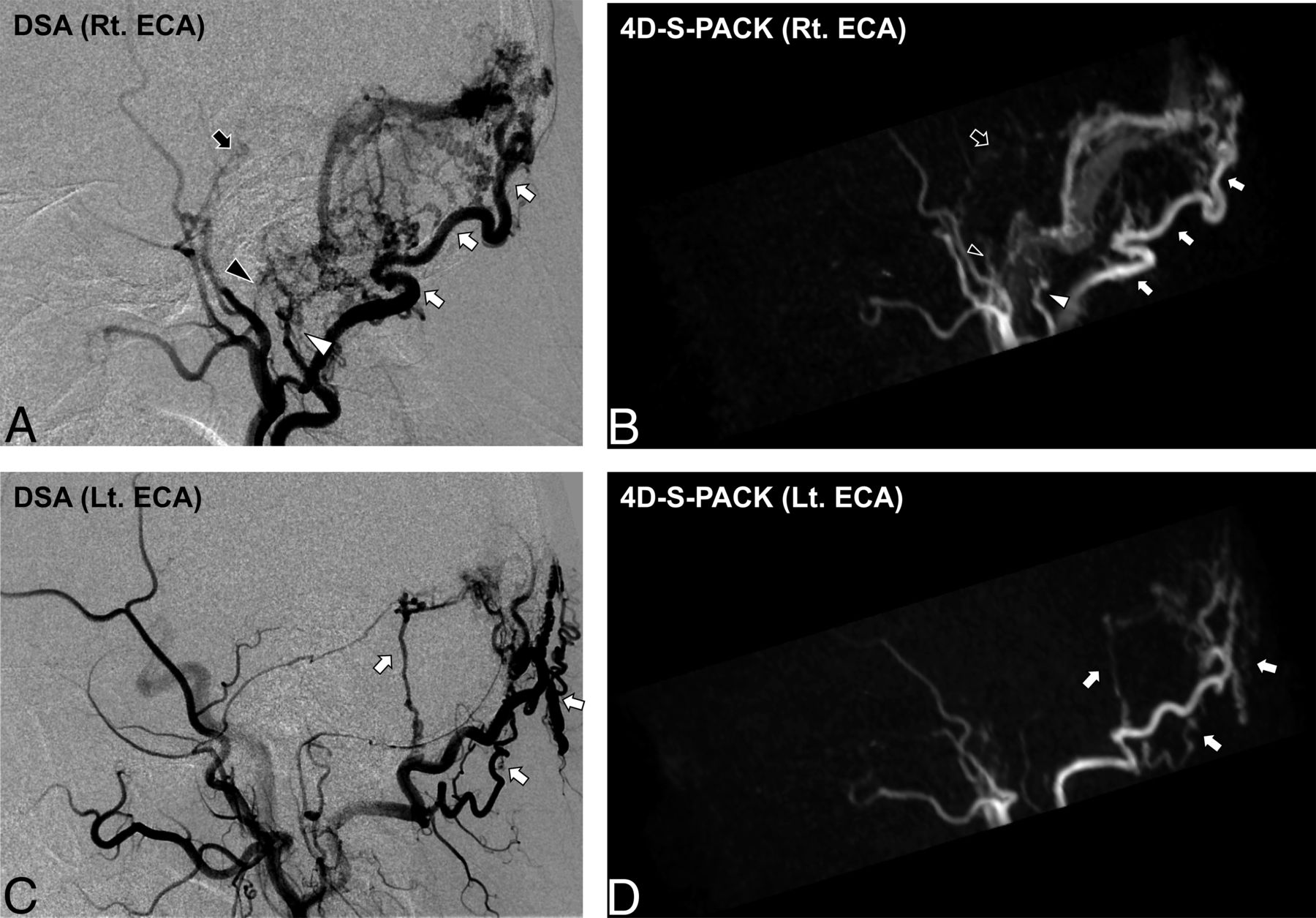
- FIG 3.
A 78-year-old man with an intracranial DAVF (Borden type I) at the transverse sigmoid sinus. DSA images (A and C) demonstrate that this shunt was supplied by multiple branches of bilateral ECAs (white arrow, occipital artery; white arrowhead, posterior auricular artery; black arrowhead, ascending pharyngeal artery; black arrow, middle meningeal artery). The 4D-S-PACK (B and D) is able to selectively image the right and left ECAs at a level comparable with that of DSA, helping to accurately identify the multiple feeding arteries. Note that no ICAs or their branches are visualized on 4D-S-PACK when the selective labeling of an ECA is performed. Rt. indicates right; Lt., left.
- FIG 4.
An 85-year-old man with an intracranial DAVF (Borden type III). DSA images (A and C) reveal that this shunt is supplied by the occipital artery branches (white arrows) at the transverse sigmoid sinus with venous drainage directly into cortical veins (black arrowheads). A varix formation (white arrowhead) is observed. In 4D-S-PACK (B and D), the left ECA is selectively imaged and the feeding artery and CVR are delineated as seen in DSA. Lt. indicates left.
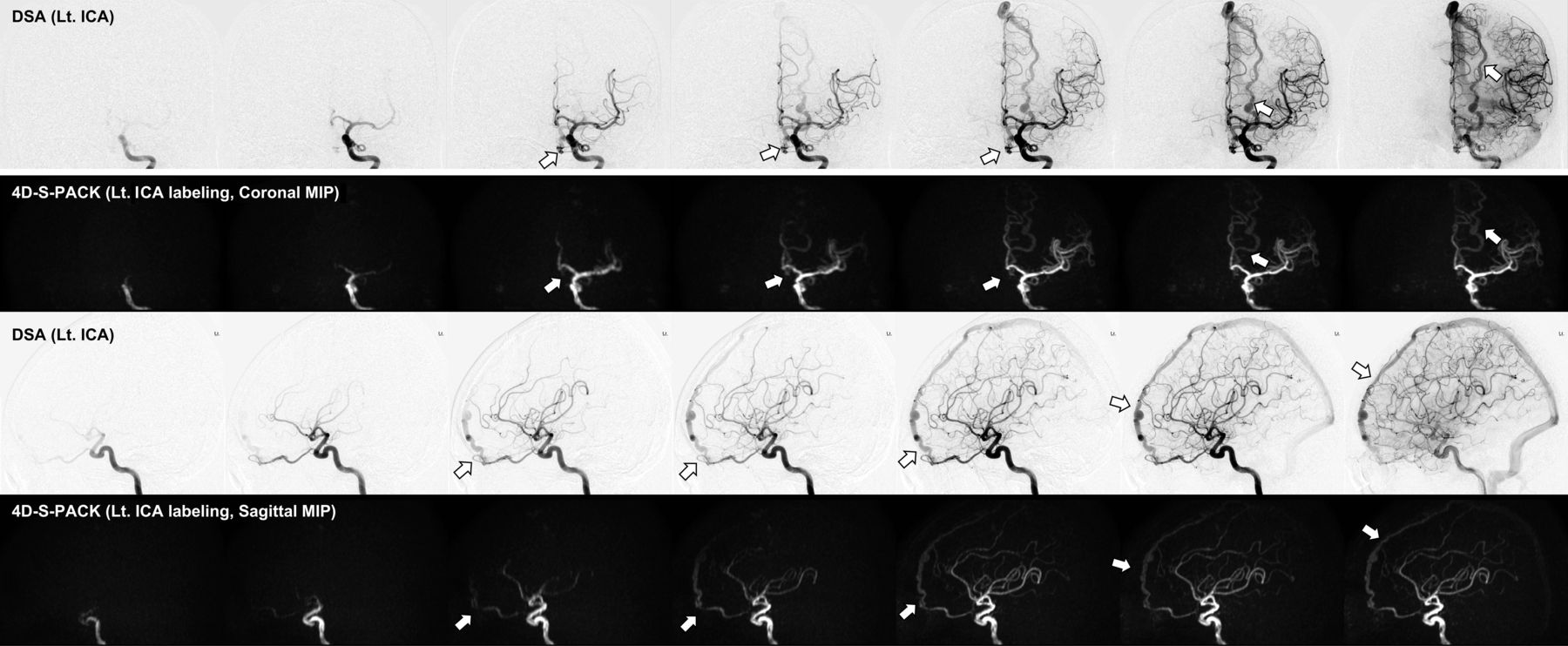
- FIG 5.
A 72-year-old woman with an intracranial DAVF (Borden type III) at the anterior skull base supplied by the left ophthalmic artery, with venous drainage directly into cortical veins and then the superior sagittal sinus (white arrow). In 4D-S-PACK, the left ICA is selectively imaged, and the feeding artery and CVR could be depicted. Lt. indicates left.


Tables
Patient No. Age Sex Shunt Location Borden Classification Symptom or Event 1 19 M R CS I Red eye 2 22 F L TS II Headache 3 27 F R CS I Visual loss, red eye 4 51 M L ASB III Free 5 51 M R CS I Headache 6 60 M R TS I Free 7 61 F L TS II Headache, tinnitus 8 62 M L TS III Loss of consciousness, intracranial hemorrhage 9 63 M R TS III Vertigo, double vision 10 65 M L TS II Tinnitus 11 70 F R TS I Tinnitus 12 71 M L CS I Headache, wamble 13 71 M R TS II Free 14 72 F L ASB III Paresthesia of the right arm 15 73 F R CS I Visual field abnormality 16 75 F L TS I Tinnitus 17 76 F L TS II Tinnitus 18 77 F R TS I Tinnitus 19 77 M SSS III Vertigo, tinnitus 20 78 M R TS I Visual loss 21 85 M L TS III Headache, intracranial hemorrhage Note:—R indicates right; L, left; TS, transverse sigmoid sinus; CS, cavernous sinus; ASB, anterior skull base; SSS, superior sagittal sinus.
Label Duration (ms) Feeding Artery Draining Vein 4D-PACK 4D-S-PACK P Value 4D-PACK 4D-S-PACK P Value 100 13.6 (SD, 12.1) 13.7 (SD, 12.6) .88 4.4 (SD, 2.6) 5.0 (SD, 3.2) .19 200 26.0 (SD, 22.3) 23.1 (SD, 18.5 .18 7.5 (SD, 6.1) 6.8 (SD, 5.1) .41 400 41.1 (SD, 26.4) 15.4 (SD, 13.0) 500 37.8 (SD, 25.9) 16.3 (SD, 14.0) 600 49.9 (SD, 24.4) 24.7 (SD, 19.6) 800 53.5 (SD, 24.6) 47.1 (SD, 24.8) .02 33.1 (SD, 23.9) 26.0 (SD, 18.7) .02 1200 64.2 (SD, 21.9) 57.2 (SD, 22.1) .03 54.7 (SD, 24.0) 39.6 (SD, 21.4) <.001 1600 66.7 (SD, 21.1) 57.8 (SD, 21.5) .006 64.2 (SD, 23.1) 45.6 (SD, 20.4) <.001 2000 59.2 (SD, 21.4) 47.3 (SD, 20.1) 2200 68.1 (SD, 21.0) 69.7 (SD, 22.4) Note:—4D-PACK indicates 4D-MR angiography based on pseudo-continuous arterial spin labeling combined with CENTRA-keyhole and view-sharing; 4D-S-PACK, 4D-MR angiography based on super-selective pCASL with CENTRA-keyhole and view-sharing.
↵a Data are expressed as mean values.


