
Article Figures & Data
Figures
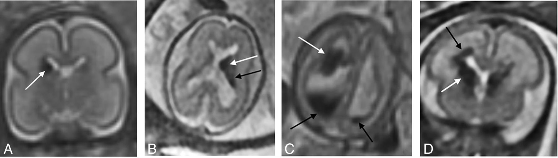
- FIG 1.
A, Grade I GMH in a 24 -weeks’ GA fetus. Coronal T2 SSFSE of the brain demonstrates T2 hypointensity at the right caudothalamic groove (white arrow). B, Grade II GMH in a 24 weeks’ GA fetus. Axial T2 SSFSE of the brain shows T2 hypointensity at the left caudothalamic groove (white arrow) extending posteriorly along the margin of the lateral ventricle (black arrow). C, Grade III GMH in a 20 weeks’ GA fetus. Axial T2 SSFSE of the brain demonstrates globular T2 hypointensity near the right caudothalamic groove (white arrow) with layering T2 hypointensity/hemorrhage in the right greater than left posterior horns of the ventricles (black arrows). There is dilation of the bilateral ventricles, right greater than left, measuring up to 16 mm on the right. D, Grade IV GMH in a 26 weeks’ GA fetus. Coronal T2 SSFSE of the brain shows T2 hypointensity at the right caudothalamic groove (white arrow), with T2 signal/hemorrhage extending into the periventricular white matter (black arrow). Grade II GMH is partially visualized on the left.
- FIG 2.
Grade 0 GMH in a 29 weeks’ GA fetus. Coronal T2 SSFSE of the brain shows T2-hyperintense cystic lesions at the bilateral caudothalamic grooves (white arrows) and abnormal T2 hyperintense signal in the bilateral basal ganglia.
- FIG 3.
Coronal T2 SSFSE (A) and axial EPI (B) of the brain in a 31 weeks’ GA fetus. Note T2 hypointensity at the margin of the inferior aspect of the deficient right cerebellar hemisphere (arrow, A) and an associated magnetic susceptibility signal at the right cerebellar hemisphere (dashed arrow, B) and vermis.
- FIG 4.
Linear regression model shows the ventricle size increased by 2.4 mm when the GMH grade increased by 1 (P < .001).
- FIG 5.
Left, grade IV GMH in a 35 weeks’ GA fetus. Axial T1(A), DWI (B), and T2 SSFSE (C) images of the brain demonstrate blood products centered in the left caudothalamic groove, extending posteriorly along the left lateral ventricle with associated T1 hyperintensity, restricted diffusion, and T2 hypointensity (arrows). D, Postnatal axial T2 of the brain at day 6 of life shows decreased T2 hypointensity centered at the left caudothalamic groove (dashed arrow, D) compared with prenatal image (C), consistent with decreased volume of hemorrhage.


Tables
Characteristics Data (n = 177) Sex Male 48.0% (85/177) Female 40.1% (71/177) Unknown 11.9% (21/177) Singleton 70.1% (124/177) Multiple gestation 29.9% (53/177) Average GA at fetal MR imaging (wk) 25.73 (SD, 5.01) GMH 60.5% (107/177) Grade 0 12.1% (13/107) Grade I 28.9% (31/107) Grade II 28.0% (30/107) Grade III 6.5% (7/107) Grade IV 24.3% (26/107) Non-GMH 39.5% (70/177) Cerebellar hemorrhages 9% (16/177) Fetal growth restriction 21.3% (20/94) Other imaging abnormalities 79.1% (140/177) Postnatal imaging 22.6% (40/177) Head US (No.) 72.5% (29/40) MR imaging (No.) 87.% (35/40) CT (No.) 52.5% (21/40) Average maternal age at fetal MR imaging (yr) 28.02 (SD, 6.02) ICH Imaging Characteristics Data (n = 177) T2 (+), n = 177 84.7% (150/177) T1 (+), n = 150 46% (69/150) DWI (+), n = 135 57.8% (78/135) EPI (+), n = 81 96.3% (78/81) Ventriculomegaly (>10 mm) 59.3% (105/177) Ventricular size (mm) 12.76 (SD, 7.54) Note:— + indicates positive imaging finding on fetal MR in those fetuses with ICH.
Imaging Modality Findings Data US Same 27.6% (8/29) Better 69.0% (20/29) Worse 3.4% (1/29) MR Same 14.3% (5/35) Better 74.3% (26/35) Worse 11.4% (4/35) CT Same 0% (0/21) Better 95.2% (20/21) Worse 4.8% (1/21)


