
Article Figures & Data
Figures
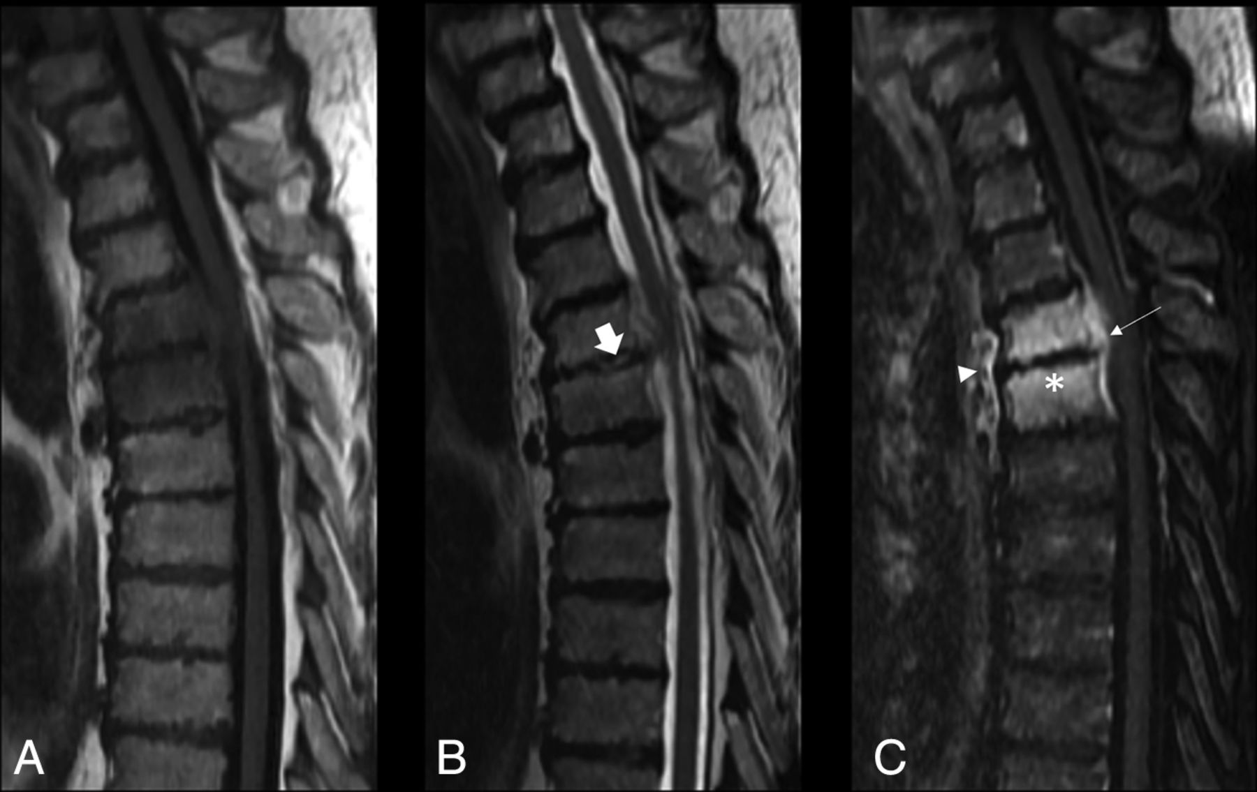
- FIG 1.
Imaging panel in a patient with osteomyelitis with hyperintense T2 disc signal, adjacent vertebral endplate erosions, and paraspinal/epidural enhancement. Thoracic spine MR imaging of a 72-year-old man with radiologically suspected infection at T4–T5. A, Disc-centered and bone marrow hypointensity on a T1-weighted image. B, Mild hyperintensity of the disc and adjacent bone marrow on T2-weighted image. The arrow represents hyperintense T2 disc signal. C, Contrast-enhanced T1-weighted image demonstrates epidural (arrow) and paraspinal enhancement (arrowhead). The asterisk represents adjacent vertebral endplate erosion. Pathology demonstrated that inflammatory histology and microbiology had no growth. ESR was 123 mm/h, and CRP was 176 mg/L. The patient was febrile on presentation with leukocytosis.

Tables
Variable No (%) Age (mean) (yr) 63 ± 16 Sex (M/F) 41:31 Immunosuppression 33 (46) Cancer 8 (11) COPD 6 (8) Cirrhosis 3 (4) Diabetes 15 (21) HIV 4 (6) Steroid use 12 (17) IV drug abuse 9 (13) Postoperative status (within 1 wk) Symptoms relevant to discitis 0 (0) Back pain 72 (100) Febrile 25 (35) Radiation 12 (17) Numbness/weakness 10 (14) Bowel or bladder incontinence 3 (4) Time to diagnosis (days)a 55 (1–270) Site of involvement Cervical 3 (4) Thoracic 20 (28) Lumbar 49 (68) Biopsy technique CT 50 (69) Fluoroscopy 22 (31) Either surgical pathology (+)/microbiology (+) 29 (40) Surgical pathology (+) 24 (33) Microbiology growth from tissue (+) 12 (17) Both 7 (10) Bacterial isolates from tissue culture 12 Staphylococci 5 (42) Streptococci 3 (25) Pseudomonas 2 (17) Klebsiella 1 (8) Mycobacteria 1 (8) Blood culture growth (+) 2 (3) Laboratory leukocyte count (cells/mm3)a 8.0 (4.1–17.5) CRP (mg/L)a 46.4 (1–303) ESR (mm/h)a 59.3 (6–156) Note:—COPD indicates chronic obstructive pulmonary disease.
↵a Mean followed by range in parenthesis.
- Table 2:
Inflammatory biomarker characteristics associated with pathology and/or microbiology for spondylodiscitis
Pathology Pathology and/or Microbiologyy Positive Negative Positive Negative CRP (mean) (mg/L) 93.4 40.2 86.4 38.3 Median 50 31.5 60.5 22.0 Range 7–303 1–156 2–303 1–156 SD 90.3 38.7 84.9 42.4 ESR (mean) (mm/h) 65.1 51.0 66.8 51.6 Median 59.0 46 61.5 38.5 Range 12–150 6–106 22–156 6–109 SD 36.5 27.5 34.9 33.3 Positive Pathologyy r P PPV NPV Sensitivity Specificity Epidural enhancement Observer 1 0.52 <.001c 61.3 87.8 79.2 75.0 Observer 2 0.36 .002c 42.1 100 100 31.3 Observer 3 0.33 .004c 40.7 100 100 27.1 Majority consensus 0.41 .001c 44.4 100 100 37.5 Paraspinal enhancement Observer 1 0.27 .02c 40.7 88.9 91.7 33.3 Observer 2 0.23 .05c 36.9 100 100 14.6 Observer 3 0.24 .04c 43.6 78.8 70.8 54.2 Majority consensus 0.33 .01c 40.7 100 100 27.1 Hyperintense T2 disc signal Observer 1 0.37 .02c 55.5 80.1 86.5 45.0 Observer 2 0.19 .03c 33.0 100 100 20.2 Observer 3 0.21 .03c 41.2 79.2 73.1 51.0 Majority consensus 0.29 .03c 51.0 100 100 33.2 Vertebral endplate erosion Observer 1 0.333 .01c 46.0 83.4 88.8 30.2 Observer 2 0.22 .05c 35.6 95.5 93.2 25.7 Observer 3 0.30 .0c 39.4 80.6 75.6 56.7 Majority consensus 0.31 .02c 42.1 92.3 92.0 35.6 Blood culture growth 0.06 .63 50 66.7 4.2 97.9 Fever status 0.26 .03c 48.3 76.7 58.3 68.8 Leukocytosis 0.032 .80 40 64.3 16.7 85.7 Hemoglobin countb –0.05 .67 NA NA NA NA Platelet countb 0.20 .11 NA NA NA NA ALP levelb –0.03 .84 NA NA NA NA ESRb –0.09 .49 NA NA NA NA CRPb 0.29 .02c NA NA NA NA Note:—NA indicates not applicable; ALP, alkaline phosphatase.
↵a The Spearman correlation was used for rank/categoric variables. PPV, NPV, sensitivity, and specificity numbers are represented in percentages. Continuous variables will not have PPV, NPV, sensitivity, or specificity values without established thresholds.
↵b Continuous variables in which the Pearson correlation was used.
↵c Statistically significant P values (<.05).
- Table 4:
Correlation between individual biomarkers and spondylodiscitis as proved on pathology and/or microbiologya
Positive Pathology and/or Microbiologyy r P PPV NPV Sensitivity Specificity Epidural enhancement Observer 1 0.37 .00b 61.3 75.6 65.5 72.1 Observer 2 0.42 .001b 50.9 100 100 34.9 Observer 3 0.39 .001b 49.2 100 100 30.2 Majority consensus 0.47 .001b 53.7 100 100 41.9 Paraspinal enhancement Observer 1 0.34 .03b 50.0 88.9 93.1 37.2 Observer 2 0.27 .02b 44.6 100 100 16.3 Observer 3 0.30 .01b 53.8 75.8 72.4 58.1 Majority consensus 0.39 .01b 49.2 100 100 30.2 Hyperintense T2 disc signal Observer 1 0.30 .03b 49.2 79.6 82.5 42.6 Observer 2 0.15 .05b 31.2 96.2 95.3 19.6 Observer 3 0.25 .02b 45.5 80.2 74.2 50.4 Majority consensus 0.28 .03b 48.0 95.2 93.5 29.8 Vertebral endplate erosion Observer 1 0.29 .02b 48.9 82.4 89.6 33.6 Observer 2 0.17 .03b 37.5 95.8 92.5 27.5 Observer 3 0.33 .01b 38.1 82.5 76.3 59.1 Majority consensus 0.31 .02b 42.6 91.5 91.8 34.7 Blood culture growth 0.03 .79 50 59.4 3.4 97.6 Fever status 0.25 .03a 55.2 69.8 55.2 69.8 Leukocytosis 0.039 .77 40 55.4 13.8 83.8 Hemoglobin countc −0.02 .86 NA NA NA NA Platelet countc 0.17 .18 NA NA NA NA ALP levelc 0.05 .73 NA NA NA NA ESRc −0.07 .62 NA NA NA NA CRPc 0.26 .04a NA NA NA NA Positive Pathologyy Positive Pathology and/or Microbiologyy AUC Sensitivity Specificity AUC Sensitivity Specificity CRP, ESR, and fever 0.72 68.2 67.0 0.68 60.5 64.5 CRP, ESR, fever, and epidural enhancement (observer 1) 0.87 83.1 79.8 0.76 68.4 75.6 CRP, ESR, fever, and epidural enhancement (observer 2) 0.76 75.0 66.5 0.73 59.6 76.2 CRP, ESR, fever, and epidural enhancement (observer 3) 0.79 77.2 70.1 0.78 69.3 75.8 CRP, ESR, fever, and epidural enhancement (majority consensus) 0.80 78.3 75.6 0.79 77.4 74.6 ↵a Logistic regression with backward stepwise selection was used to find the optimal combination of clinical and imaging features for 3 independent observers with majority consensus among the observers.


