Advertisement

Main menu

  • Home
  • Content
    • Current Issue
    • Accepted Manuscripts
    • Article Preview
    • Past Issue Archive
    • Video Articles
    • AJNR Case Collection
    • Case of the Week Archive
    • Case of the Month Archive
    • Classic Case Archive
  • Special Collections
    • AJNR Awards
    • Low-Field MRI
    • Alzheimer Disease
    • ASNR Foundation Special Collection
    • Photon-Counting CT
    • View All
  • Multimedia
    • AJNR Podcasts
    • AJNR SCANtastic
    • Trainee Corner
    • MRI Safety Corner
    • Imaging Protocols
  • For Authors
    • Submit a Manuscript
    • Submit a Video Article
    • Submit an eLetter to the Editor/Response
    • Manuscript Submission Guidelines
    • Statistical Tips
    • Fast Publishing of Accepted Manuscripts
    • Graphical Abstract Preparation
    • Imaging Protocol Submission
    • Author Policies
  • About Us
    • About AJNR
    • Editorial Board
    • Editorial Board Alumni
  • More
    • Become a Reviewer/Academy of Reviewers
    • Subscribers
    • Permissions
    • Alerts
    • Feedback
    • Advertisers
    • ASNR Home

User menu

  • Alerts
  • Log in

Search

  • Advanced search
American Journal of Neuroradiology

American Journal of Neuroradiology

ASHNR American Society of Functional Neuroradiology ASHNR American Society of Pediatric Neuroradiology ASSR
  • Alerts
  • Log in

Advanced Search

  • Home
  • Content
    • Current Issue
    • Accepted Manuscripts
    • Article Preview
    • Past Issue Archive
    • Video Articles
    • AJNR Case Collection
    • Case of the Week Archive
    • Case of the Month Archive
    • Classic Case Archive
  • Special Collections
    • AJNR Awards
    • Low-Field MRI
    • Alzheimer Disease
    • ASNR Foundation Special Collection
    • Photon-Counting CT
    • View All
  • Multimedia
    • AJNR Podcasts
    • AJNR SCANtastic
    • Trainee Corner
    • MRI Safety Corner
    • Imaging Protocols
  • For Authors
    • Submit a Manuscript
    • Submit a Video Article
    • Submit an eLetter to the Editor/Response
    • Manuscript Submission Guidelines
    • Statistical Tips
    • Fast Publishing of Accepted Manuscripts
    • Graphical Abstract Preparation
    • Imaging Protocol Submission
    • Author Policies
  • About Us
    • About AJNR
    • Editorial Board
    • Editorial Board Alumni
  • More
    • Become a Reviewer/Academy of Reviewers
    • Subscribers
    • Permissions
    • Alerts
    • Feedback
    • Advertisers
    • ASNR Home
  • Follow AJNR on Twitter
  • Visit AJNR on Facebook
  • Follow AJNR on Instagram
  • Join AJNR on LinkedIn
  • RSS Feeds

AJNR Awards, New Junior Editors, and more. Read the latest AJNR updates

Getting new auth cookie, if you see this message a lot, tell someone!
Research ArticleSpine

Decubitus CT Myelography for Detecting Subtle CSF Leaks in Spontaneous Intracranial Hypotension

P.G. Kranz, L. Gray and T.J. Amrhein
American Journal of Neuroradiology April 2019, 40 (4) 754-756; DOI: https://doi.org/10.3174/ajnr.A5995
P.G. Kranz
aFrom the Department of Radiology, Duke University Medical Center, Durham, North Carolina.
  • Find this author on Google Scholar
  • Find this author on PubMed
  • Search for this author on this site
  • ORCID record for P.G. Kranz
L. Gray
aFrom the Department of Radiology, Duke University Medical Center, Durham, North Carolina.
  • Find this author on Google Scholar
  • Find this author on PubMed
  • Search for this author on this site
  • ORCID record for L. Gray
T.J. Amrhein
aFrom the Department of Radiology, Duke University Medical Center, Durham, North Carolina.
  • Find this author on Google Scholar
  • Find this author on PubMed
  • Search for this author on this site
  • ORCID record for T.J. Amrhein
  • Article
  • Figures & Data
  • Info & Metrics
  • Responses
  • References
  • PDF
Loading

Article Figures & Data

Figures

  • Fig 1.
    • Download figure
    • Open in new tab
    • Download powerpoint
    Fig 1.

    CSF venous fistula visualized best on decubitus CTM. Axial prone CTM image (A) and maximum-intensity-projection image of the same level (B) show subtle linear contrast (arrows) lateral to the T11 nerve root. The patient was turned to the left lateral decubitus position and re-scanned 13 minutes later. Axial MIP image (C) from that scan shows increased intravascular contrast with the patient in the decubitus position, suggestive of a CSF venous fistula (arrow). Axial MIP image (D) from a CTM performed after dynamic myelography on a subsequent day with the patient maintained in the decubitus position after contrast injection shows extensive filling of a paraspinal vein distal to the fistula (arrows).
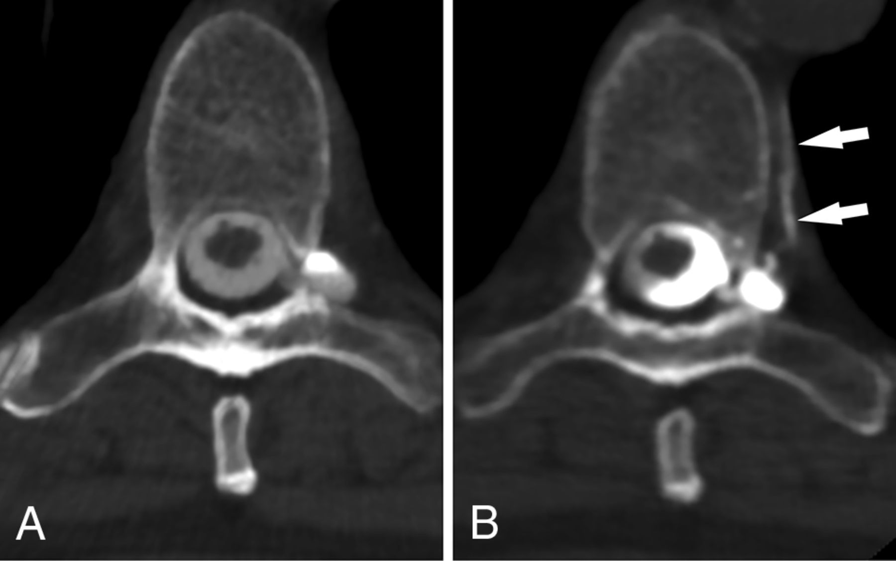
  • Fig 2.
    • Download figure
    • Open in new tab
    • Download powerpoint
    Fig 2.

    CSF venous fistula visualized best on decubitus CTM. Axial prone CT myelogram (A) shows subtle filling of a network of paraspinal veins (arrow) lateral to the nerve root and in the adjacent spinal segmental vein (arrowhead). Axial image (B) from a decubitus CTM performed after dynamic myelography on a subsequent day shows increased filling of the lateral veins (arrow) and segmental vein (arrowheads), making the CVF much more conspicuous.
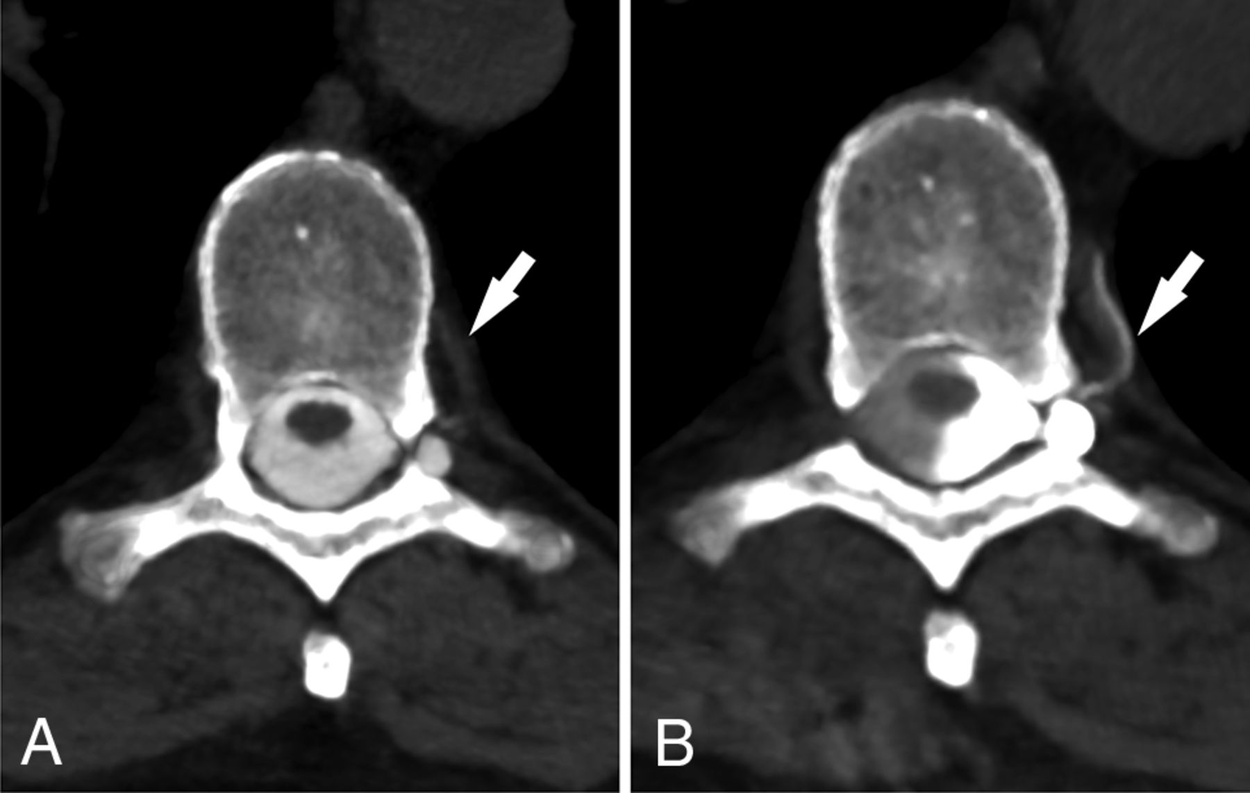
  • Fig 3.
    • Download figure
    • Open in new tab
    • Download powerpoint
    Fig 3.

    CSF venous fistula visualized best on decubitus CTM. Axial prone CTM image (A) shows possible increased density of a spinal segmental vein (arrow). Axial image (B) from a decubitus CTM performed after dynamic myelography on a subsequent day shows increased filling of the segmental veins (arrow), helping to confirm the diagnosis of CVF. Note the increased density of the left lateral thecal sac due to decubitus positioning.

  • Fig 4.
    • Download figure
    • Open in new tab
    • Download powerpoint
    Fig 4.

    CSF venous fistula visualized best on decubitus CTM. Axial prone CTM image (A) shows a perineural diverticulum, but no clear leak. Axial image (B) from a subsequent CTM obtained with the patient in the decubitus position after dynamic myelography shows clear filling of a segmental vein (arrows), confirming a CVF.
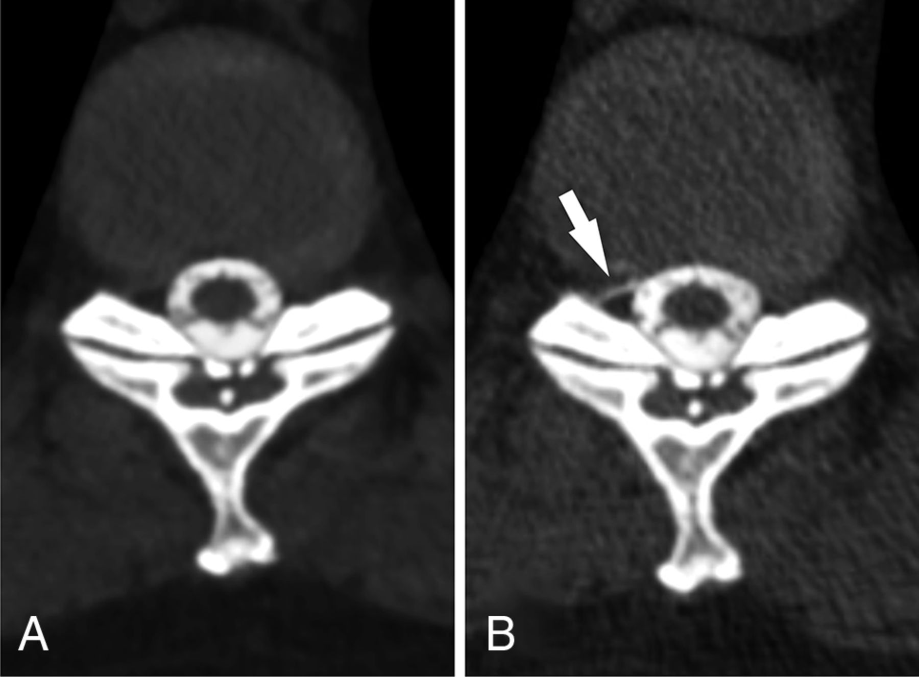
  • Fig 5.
    • Download figure
    • Open in new tab
    • Download powerpoint
    Fig 5.

    Low-flow CSF leak seen only on decubitus myelography. Axial prone CTM image (A) shows no CSF leak at the T10–11 level. The patient was turned into the left lateral decubitus position and re-scanned, again with the scan showing no leak (not shown). The patient was then turned to the right lateral decubitus position. An axial CTM image from the decubitus myelogram (B) obtained 6 minutes later shows a low-flow CSF leak not seen on prone myelogram (arrow).

PreviousNext
Back to top

In this issue

American Journal of Neuroradiology: 40 (4)
American Journal of Neuroradiology
Vol. 40, Issue 4
1 Apr 2019
  • Table of Contents
  • Index by author
  • Complete Issue (PDF)
Advertisement
Print
Download PDF
Email Article

Thank you for your interest in spreading the word on American Journal of Neuroradiology.

NOTE: We only request your email address so that the person you are recommending the page to knows that you wanted them to see it, and that it is not junk mail. We do not capture any email address.

Enter multiple addresses on separate lines or separate them with commas.
Decubitus CT Myelography for Detecting Subtle CSF Leaks in Spontaneous Intracranial Hypotension
(Your Name) has sent you a message from American Journal of Neuroradiology
(Your Name) thought you would like to see the American Journal of Neuroradiology web site.
CAPTCHA
This question is for testing whether or not you are a human visitor and to prevent automated spam submissions.
Cite this article
P.G. Kranz, L. Gray, T.J. Amrhein
Decubitus CT Myelography for Detecting Subtle CSF Leaks in Spontaneous Intracranial Hypotension
American Journal of Neuroradiology Apr 2019, 40 (4) 754-756; DOI: 10.3174/ajnr.A5995

Citation Manager Formats

  • BibTeX
  • Bookends
  • EasyBib
  • EndNote (tagged)
  • EndNote 8 (xml)
  • Medlars
  • Mendeley
  • Papers
  • RefWorks Tagged
  • Ref Manager
  • RIS
  • Zotero
0 Responses
Respond to this article
Share
Bookmark this article
Decubitus CT Myelography for Detecting Subtle CSF Leaks in Spontaneous Intracranial Hypotension
P.G. Kranz, L. Gray, T.J. Amrhein
American Journal of Neuroradiology Apr 2019, 40 (4) 754-756; DOI: 10.3174/ajnr.A5995
del.icio.us logo Twitter logo Facebook logo Mendeley logo
  • Tweet Widget
  • Facebook Like
  • Google Plus One
Purchase

Jump to section

  • Article
    • Abstract
    • ABBREVIATIONS:
    • Materials and Methods
    • Results
    • Discussion
    • Conclusions
    • Footnotes
    • REFERENCES
  • Figures & Data
  • Info & Metrics
  • Responses
  • References
  • PDF

Related Articles

  • No related articles found.
  • PubMed
  • Google Scholar

Cited By...

  • Density and Time Characteristics of CSF-Venous Fistulas on CT Myelography in Patients with Spontaneous Intracranial Hypotension
  • CSF-Venous Fistulas Arising Intraosseously within Bone Remodeled by Meningeal Diverticula
  • Safety and Technical Performance of Bilateral Decubitus CT Myelography Using Standard versus Increased Intrathecal Iodinated Contrast Volume
  • Spontaneous Intracranial Hypotension in Children: A Multi-Institutional Review of Spinal CSF Leaks Localized on Advanced Myelography
  • Direct comparison of digital subtraction myelography versus CT myelography in lateral decubitus position: evaluation of diagnostic yield for cerebrospinal fluid-venous fistulas
  • Diagnostic Yield of Decubitus CT Myelography for Detection of CSF-Venous Fistulas
  • Myelographic Techniques for the Localization of CSF-Venous Fistulas: Updates in 2024
  • Lateral Decubitus Dynamic CT Myelography with Real-Time Bolus Tracking (dCTM-BT) for Evaluation of CSF-Venous Fistulas: Diagnostic Yield Stratified by Brain Imaging Findings
  • Temporal Characteristics of CSF-Venous Fistulas on Dynamic Decubitus CT Myelography: A Retrospective Multi-Institution Cohort Study
  • Diagnostic Performance of Decubitus Photon-Counting Detector CT Myelography for the Detection of CSF-Venous Fistulas
  • A Novel Patient-Positioning Device for Dynamic CT Myelography
  • Resisted Inspiration Improves Visualization of CSF-Venous Fistulas in Spontaneous Intracranial Hypotension
  • Utility of Photon-Counting Detector CT Myelography for the Detection of CSF-Venous Fistulas
  • Utility of Photon-Counting Detector CT Myelography for the Detection of CSF-Venous Fistulas
  • Conebeam CT as an Adjunct to Digital Subtraction Myelography for Detection of CSF-Venous Fistulas
  • The promise and challenges of CSF-venous fistula treatment
  • Surgical Ligation of Spinal CSF-Venous Fistulas after Transvenous Embolization in Patients with Spontaneous Intracranial Hypotension
  • Same-Day Bilateral Decubitus CT Myelography for Detecting CSF-Venous Fistulas in Spontaneous Intracranial Hypotension
  • Spinal Compliance Curves: Preliminary Experience with a New Tool for Evaluating Suspected CSF Venous Fistulas on CT Myelography in Patients with Spontaneous Intracranial Hypotension
  • A Novel Endovascular Therapy for CSF Hypotension Secondary to CSF-Venous Fistulas
  • Decubitus CT Myelography for CSF-Venous Fistulas: A Procedural Approach
  • Respiratory Phase Affects the Conspicuity of CSF-Venous Fistulas in Spontaneous Intracranial Hypotension
  • MR Myelography for the Detection of CSF-Venous Fistulas
  • Crossref
  • Google Scholar

This article has not yet been cited by articles in journals that are participating in Crossref Cited-by Linking.

More in this TOC Section

  • CT-Guided C2 Dorsal Root Ganglion Radiofrequency Ablation for the Treatment of Cervicogenic Headache: Case Series and Clinical Outcomes
  • The Evaluation and Prediction of Laminoplasty Surgery Outcome in Patients with Degenerative Cervical Myelopathy Using Diffusion Tensor MRI
  • Subject-Specific Studies of CSF Bulk Flow Patterns in the Spinal Canal: Implications for the Dispersion of Solute Particles in Intrathecal Drug Delivery
Show more Spine

Similar Articles

Advertisement

Indexed Content

  • Current Issue
  • Accepted Manuscripts
  • Article Preview
  • Past Issues
  • Editorials
  • Editor's Choice
  • Fellows' Journal Club
  • Letters to the Editor
  • Video Articles

Cases

  • Case Collection
  • Archive - Case of the Week
  • Archive - Case of the Month
  • Archive - Classic Case

More from AJNR

  • Trainee Corner
  • Imaging Protocols
  • MRI Safety Corner
  • Book Reviews

Multimedia

  • AJNR Podcasts
  • AJNR Scantastics

Resources

  • Turnaround Time
  • Submit a Manuscript
  • Submit a Video Article
  • Submit an eLetter to the Editor/Response
  • Manuscript Submission Guidelines
  • Statistical Tips
  • Fast Publishing of Accepted Manuscripts
  • Graphical Abstract Preparation
  • Imaging Protocol Submission
  • Evidence-Based Medicine Level Guide
  • Publishing Checklists
  • Author Policies
  • Become a Reviewer/Academy of Reviewers
  • News and Updates

About Us

  • About AJNR
  • Editorial Board
  • Editorial Board Alumni
  • Alerts
  • Permissions
  • Not an AJNR Subscriber? Join Now
  • Advertise with Us
  • Librarian Resources
  • Feedback
  • Terms and Conditions
  • AJNR Editorial Board Alumni

American Society of Neuroradiology

  • Not an ASNR Member? Join Now

© 2025 by the American Society of Neuroradiology All rights, including for text and data mining, AI training, and similar technologies, are reserved.
Print ISSN: 0195-6108 Online ISSN: 1936-959X

Powered by HighWire