Advertisement

Main menu

  • Home
  • Content
    • Current Issue
    • Accepted Manuscripts
    • Article Preview
    • Past Issue Archive
    • Video Articles
    • AJNR Case Collection
    • Case of the Week Archive
    • Case of the Month Archive
    • Classic Case Archive
  • Special Collections
    • AJNR Awards
    • Low-Field MRI
    • Alzheimer Disease
    • ASNR Foundation Special Collection
    • Photon-Counting CT
    • View All
  • Multimedia
    • AJNR Podcasts
    • AJNR SCANtastic
    • Trainee Corner
    • MRI Safety Corner
    • Imaging Protocols
  • For Authors
    • Submit a Manuscript
    • Submit a Video Article
    • Submit an eLetter to the Editor/Response
    • Manuscript Submission Guidelines
    • Statistical Tips
    • Fast Publishing of Accepted Manuscripts
    • Graphical Abstract Preparation
    • Imaging Protocol Submission
    • Author Policies
  • About Us
    • About AJNR
    • Editorial Board
    • Editorial Board Alumni
  • More
    • Become a Reviewer/Academy of Reviewers
    • Subscribers
    • Permissions
    • Alerts
    • Feedback
    • Advertisers
    • ASNR Home

User menu

  • Alerts
  • Log in

Search

  • Advanced search
American Journal of Neuroradiology

American Journal of Neuroradiology

ASHNR American Society of Functional Neuroradiology ASHNR American Society of Pediatric Neuroradiology ASSR
  • Alerts
  • Log in

Advanced Search

  • Home
  • Content
    • Current Issue
    • Accepted Manuscripts
    • Article Preview
    • Past Issue Archive
    • Video Articles
    • AJNR Case Collection
    • Case of the Week Archive
    • Case of the Month Archive
    • Classic Case Archive
  • Special Collections
    • AJNR Awards
    • Low-Field MRI
    • Alzheimer Disease
    • ASNR Foundation Special Collection
    • Photon-Counting CT
    • View All
  • Multimedia
    • AJNR Podcasts
    • AJNR SCANtastic
    • Trainee Corner
    • MRI Safety Corner
    • Imaging Protocols
  • For Authors
    • Submit a Manuscript
    • Submit a Video Article
    • Submit an eLetter to the Editor/Response
    • Manuscript Submission Guidelines
    • Statistical Tips
    • Fast Publishing of Accepted Manuscripts
    • Graphical Abstract Preparation
    • Imaging Protocol Submission
    • Author Policies
  • About Us
    • About AJNR
    • Editorial Board
    • Editorial Board Alumni
  • More
    • Become a Reviewer/Academy of Reviewers
    • Subscribers
    • Permissions
    • Alerts
    • Feedback
    • Advertisers
    • ASNR Home
  • Follow AJNR on Twitter
  • Visit AJNR on Facebook
  • Follow AJNR on Instagram
  • Join AJNR on LinkedIn
  • RSS Feeds

AJNR Awards, New Junior Editors, and more. Read the latest AJNR updates

Letter

Comment on “Aneurysms Associated with Brain Arteriovenous Malformations”

F. Clarençon, E. Shotar and N.-A. Sourour
American Journal of Neuroradiology January 2017, 38 (1) E1-E4; DOI: https://doi.org/10.3174/ajnr.A4966
F. Clarençon
aDepartment of Interventional Neuroradiology Pitié-Salpêtrière Hospital AP-HP Paris, France
bParis VI University (Pierre et Marie Curie University) Paris, France
  • Find this author on Google Scholar
  • Find this author on PubMed
  • Search for this author on this site
  • ORCID record for F. Clarençon
E. Shotar
aDepartment of Interventional Neuroradiology Pitié-Salpêtrière Hospital AP-HP Paris, France
bParis VI University (Pierre et Marie Curie University) Paris, France
  • Find this author on Google Scholar
  • Find this author on PubMed
  • Search for this author on this site
  • ORCID record for E. Shotar
N.-A. Sourour
cDepartment of Interventional Neuroradiology Pitié-Salpêtrière Hospital AP-HP Paris, France
  • Find this author on Google Scholar
  • Find this author on PubMed
  • Search for this author on this site
  • ORCID record for N.-A. Sourour
  • Article
  • Figures & Data
  • Info & Metrics
  • Responses
  • References
  • PDF
Loading

We read with great interest the review article entitled “Aneurysms Associated with Brain Arteriovenous Malformations” by Rammos et al.1 This article summarizes the different subtypes of intracranial aneurysms that may be associated with brain AVMs (bAVMs). We congratulate the authors for their effort to clarify the nomenclature of such aneurysms. However, we would like to raise our disagreement with the following statement: “Given that pathologic specimens of resected AVM nidi consist of a conglomerate of venous tangles and loops, implicating that venous drainage begins at the level of the nidus, intranidal aneurysms are de facto venous.” Histopathologically, the nidus has been reported to comprise both arteries and veins with disorganized walls, as well as vessels of ambiguous nature.2⇓⇓–5 Even though we agree that some intranidal aneurysms may have a venous nature and may be responsible for some bAVM bleeding events (Fig 1), in our experience, most intranidal aneurysms in bAVMs with hemorrhagic presentation are rather false aneurysms arising from the dysplastic vessels belonging to the nidus and represent the bleeding site.6 Indeed, as reported by numerous series,7,8 in patients with intracranial bAVM-related bleeding events, intranidal aneurysms are frequently observed. A higher rebleeding rate has also been reported in patients with bAVMs harboring intranidal aneurysms.9 Nidal aneurysms have been defined by a joint experts group as those with any portion contained in the bAVM nidus.10 As underlined by Redekop et al,8 these aneurysms are often seen on early phase of the DSA, before substantial venous filling, and may present a contrast media stagnation on late phase (Figs 2 and 3). Depiction of intranidal aneurysms is both difficult and subject to a high rate of interobserver disagreement. The review of Rammos et al1 is lacking a discussion on advanced imaging tools that may help to demonstrate these aneurysms and further our understanding of their origin. Indeed, high-rate (6 frames per second or more) DSA acquisitions, 3D rotational angiography (RA), 4D-RA technology,11 and ultraselective angiography, or even more recent algorithms used on 3D-RA such as segmentation algorithms12 or anamorphosis algorithm,13 may help to depict these aneurysms more accurately. Wall enhancement of the intranidal aneurysm on MR imaging has also been proposed as a radiologic feature that may help to confirm the bleeding site.14

Fig 1.
  • Download figure
  • Open in new tab
  • Download powerpoint
Fig 1.

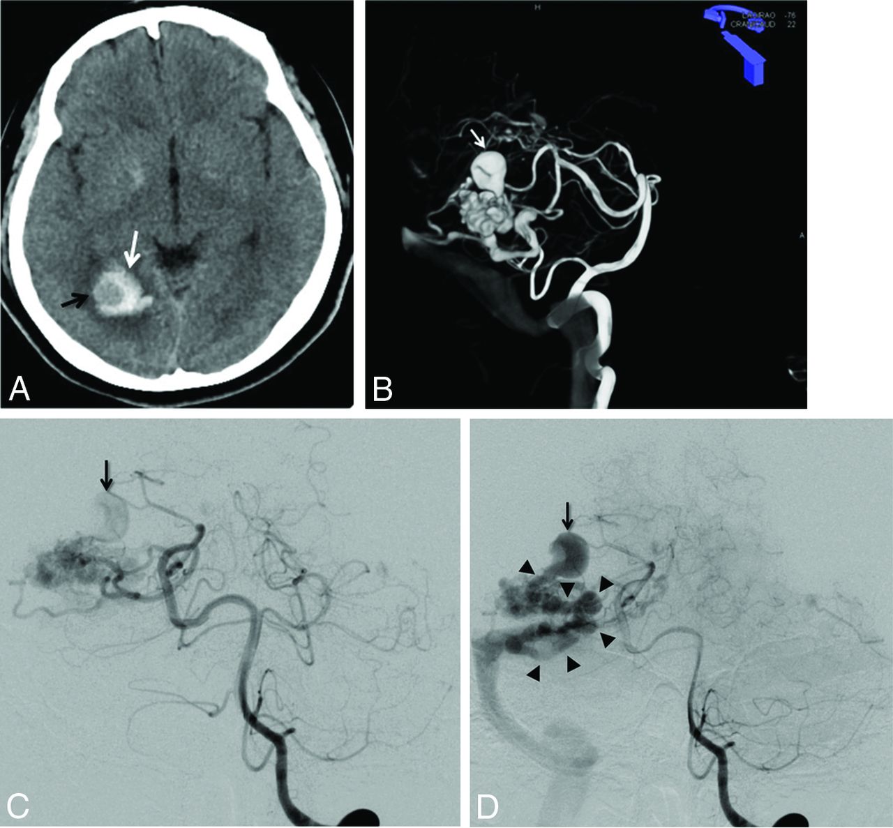
A, Unenhanced brain CT scan (axial section) in a 40-year-old woman with headache. Right occipital hematoma is seen (white arrow). Note a round hypoattenuated shape surrounded by the hematoma, corresponding to the intranidal aneurysm (black arrow). B, Volume rendering reconstruction from the 3D-RA acquisition through the left vertebral artery, showing a large intranidal aneurysm (white arrow). C and D, Left vertebral DSA in anteroposterior projection at early phase (C) and late phase (D). At early phase, an intranidal aneurysm is seen (C, arrow). On late phase, this nidal aneurysm appears clearly connected to the main draining vein (D, arrowheads), confirming the venous nature of this intranidal aneurysm.

Fig 2.
  • Download figure
  • Open in new tab
  • Download powerpoint
Fig 2.

A and B, Left vertebral artery DSA in lateral projection in a 41-year-old woman with a ruptured right parieto-occipital bAVM. A, Early phase showing a nidal aneurysm (black arrow) before filling of the draining vein. B, Intermediate phase showing a second intranidal aneurysm (white arrow) in addition to the first one (black arrow). Note a focal venous ectasia on the main draining vein (white dotted arrow). C and D, Selective DSA in anteroposterior projection from the parieto-occipital branch of the right posterior cerebral artery through a flow-dependent microcatheter. C, Early phase showing the filling of both the nidus and an intranidal aneurysm before any venous filling. D, Intermediate phase showing, in addition to the previously described intranidal aneurysm (black arrow), a second smaller nidal aneurysm (double arrow) close to the origin of the main draining vein (arrowheads). Note the presence of a focal venous ectasia on the main draining vein (black dotted arrow).

Fig 3.
  • Download figure
  • Open in new tab
  • Download powerpoint
Fig 3.

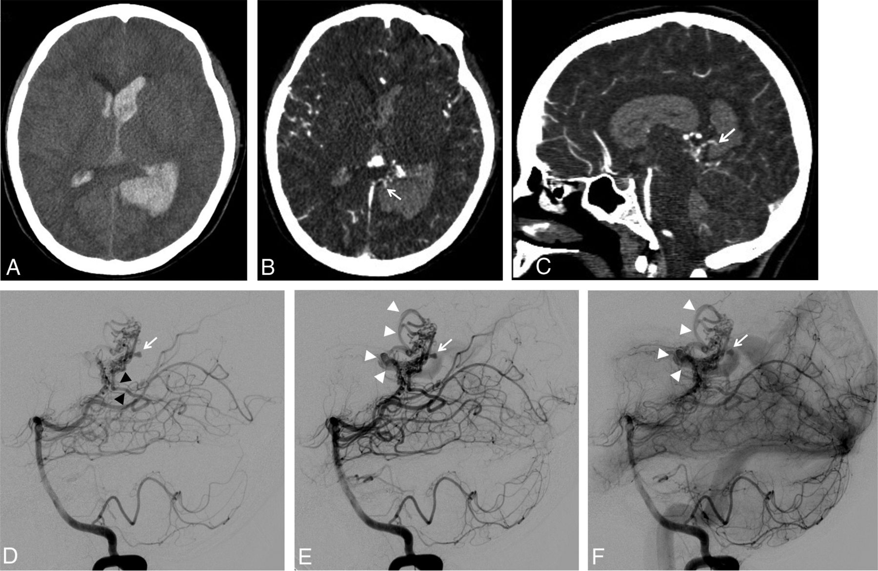
A, Unenhanced brain CT scan (axial section) showing a left parieto-occipital hematoma associated with intraventricular hemorrhage in a 19-year-old woman. B and C, Brain CT angiography: axial section (B) and sagittal reconstruction (C). Small false aneurysm arising from the nidus and close to the hematoma is seen on both axial and sagittal images (B and C, white arrows). The close relationship between the intranidal aneurysm and the hematoma suggests the aneurysm as being the cause of the bleeding. D–F, Left vertebral artery DSA in lateral projection at very early phase (D), early phase (E), and intermediate phase (F). At very early phase, opacification of the nidus is seen, supplied mainly by the left posterolateral choroidal artery (D, black arrowheads). Note the opacification of an intranidal aneurysm located at the posterior aspect of the nidus (D, white arrow) before any substantial filling of the venous drainage. At later phase, the origin of the draining vein is filling (E, white arrowheads) while the nidal aneurysm is still visible (E, white arrow). On intermediate phase, stagnation of the nidal aneurysm is seen (F, white arrow) while the venous drainage is more clearly seen (F, white arrowheads).

Finally, we agree with Rammos et al1 on the fact that the depiction of these intranidal aneurysms is of tremendous importance because they may prompt the interventional neuroradiologists or neurosurgeons to perform in an emergency a target treatment or whole resection of the bAVM to prevent early rebleeding.9

Footnotes

  • Disclosures: Frédéric Clarençon—UNRELATED: Payment for lectures including service on speakers bureaus: Medtronic. Nader Sourour—UNRELATED: Consultancy: Medtronic, Microvention; Stock/stock options: Medina/Medtronic.

References

  1. 1.↵
    1. Rammos SK,
    2. Gardenghi B,
    3. Bortolotti C, et al
    . Aneurysms associated with brain arteriovenous malformations. AJNR Am J Neuroradiol 2016 Jun 23. [Epub ahead of print] doi:10.3174/ajnr.A4869 pmid:27339951
    CrossRefPubMed
  2. 2.↵
    1. McCormick WF
    . The pathology of vascular (“arteriovenous”) malformations. J Neurosurg 1966;24:807–16 doi:10.3171/jns.1966.24.4.0807 pmid:5934138
    CrossRefPubMed
  3. 3.↵
    1. Mandybur TI,
    2. Nazek M
    . Cerebral arteriovenous malformations. A detailed morphological and immunohistochemical study using actin. Arch Pathol Lab Med 1990;114:970–73 pmid:1697158
    PubMed
  4. 4.↵
    1. Jellinger K
    . Vascular malformations of the central nervous system: a morphological overview. Neurosurg Rev 1986;9:177–216 pmid:3550522
    CrossRefPubMed
  5. 5.↵
    1. Deshpande DH,
    2. Vidyasagar C
    . Histology of the persistent embryonic veins in arteriovenous malformations of brain. Acta Neurochir (Wien) 1980;53:227–36 pmid:7424616
    CrossRefPubMed
  6. 6.↵
    1. Mjoli N,
    2. Le Feuvre D,
    3. Taylor A
    . Bleeding source identification and treatment in brain arteriovenous malformations. Interv Neuroradiol 2011;17:323–30 pmid:22005694
    Abstract/FREE Full Text
  7. 7.↵
    1. Turjman F,
    2. Massoud TF,
    3. Viñuela F, et al
    . Correlation of the angioarchitectural features of cerebral arteriovenous malformations with clinical presentation of hemorrhage. Neurosurgery 1995;37:856–60; discussion 860–62 pmid:8559332
    CrossRefPubMed
  8. 8.↵
    1. Redekop G,
    2. TerBrugge K,
    3. Montanera W, et al
    . Arterial aneurysms associated with cerebral arteriovenous malformations: classification, incidence, and risk of hemorrhage. J Neurosurg 1998;89:539–46 doi:10.3171/jns.1998.89.4.0539 pmid:9761046
    CrossRefPubMed
  9. 9.↵
    1. Meisel HJ,
    2. Mansmann U,
    3. Alvarez H, et al
    . Cerebral arteriovenous malformations and associated aneurysms: analysis of 305 cases from a series of 662 patients. Neurosurgery 2000;46:793–800; discussion 800–02 pmid:10764251
    PubMed
  10. 10.↵
    1. Atkinson RP,
    2. Awad IA,
    3. Batjer HH, et al
    . Reporting terminology for brain arteriovenous malformation clinical and radiographic features for use in clinical trials. Stroke 2001;32:1430–42 pmid:11387510
    Abstract/FREE Full Text
  11. 11.↵
    1. Lescher S,
    2. Gehrisch S,
    3. Klein S, et al
    . Time-resolved 3D rotational angiography: display of detailed neurovascular anatomy in patients with intracranial vascular malformations. J Neurointerv Surg 2016 Aug 4. [Epub ahead of print] doi:10.1136/neurintsurg-2016-012462 pmid:27492375
    Abstract/FREE Full Text
  12. 12.↵
    1. Clarençon F,
    2. Maizeroi-Eugène F,
    3. Bresson D, et al
    . Elaboration of a semi-automated algorithm for brain arteriovenous malformation segmentation: initial results. Eur Radiol 2015;25:436–43 doi:10.1007/s00330-014-3421-5 pmid:25239185
    CrossRefPubMed
  13. 13.↵
    1. Clarençon F,
    2. Maizeroi-Eugène F,
    3. Maingreaud F, et al
    . Interest of convex spherical anamorphosis in better understanding of brain AVMs' angioarchitecture. J Neurointerv Surg 2016;8:959–64 doi:10.1136/neurintsurg-2015-011888 pmid:26362085
    Abstract/FREE Full Text
  14. 14.↵
    1. Omodaka S,
    2. Endo H,
    3. Fujimura M, et al
    . High-grade cerebral arteriovenous malformation treated with targeted embolization of a ruptured site: wall enhancement of an intranidal aneurysm as a sign of ruptured site. Neurol Med Chir (Tokyo) 2015;55:813–17 doi:10.2176/nmc.cr.2015-0052 pmid:26369871
    CrossRefPubMed
  • © 2017 by American Journal of Neuroradiology
PreviousNext
Back to top

In this issue

American Journal of Neuroradiology: 38 (1)
American Journal of Neuroradiology
Vol. 38, Issue 1
1 Jan 2017
  • Table of Contents
  • Index by author
  • Complete Issue (PDF)
Advertisement
Print
Download PDF
Email Article

Thank you for your interest in spreading the word on American Journal of Neuroradiology.

NOTE: We only request your email address so that the person you are recommending the page to knows that you wanted them to see it, and that it is not junk mail. We do not capture any email address.

Enter multiple addresses on separate lines or separate them with commas.
Comment on “Aneurysms Associated with Brain Arteriovenous Malformations”
(Your Name) has sent you a message from American Journal of Neuroradiology
(Your Name) thought you would like to see the American Journal of Neuroradiology web site.
CAPTCHA
This question is for testing whether or not you are a human visitor and to prevent automated spam submissions.
Cite this article
F. Clarençon, E. Shotar, N.-A. Sourour
Comment on “Aneurysms Associated with Brain Arteriovenous Malformations”
American Journal of Neuroradiology Jan 2017, 38 (1) E1-E4; DOI: 10.3174/ajnr.A4966

Citation Manager Formats

  • BibTeX
  • Bookends
  • EasyBib
  • EndNote (tagged)
  • EndNote 8 (xml)
  • Medlars
  • Mendeley
  • Papers
  • RefWorks Tagged
  • Ref Manager
  • RIS
  • Zotero
0 Responses
Respond to this article
Share
Bookmark this article
Comment on “Aneurysms Associated with Brain Arteriovenous Malformations”
F. Clarençon, E. Shotar, N.-A. Sourour
American Journal of Neuroradiology Jan 2017, 38 (1) E1-E4; DOI: 10.3174/ajnr.A4966
del.icio.us logo Twitter logo Facebook logo Mendeley logo
  • Tweet Widget
  • Facebook Like
  • Google Plus One
Purchase

Jump to section

  • Article
    • Footnotes
    • References
  • Figures & Data
  • Info & Metrics
  • Responses
  • References
  • PDF

Related Articles

  • Reply:
  • PubMed
  • Google Scholar

Cited By...

  • No citing articles found.
  • Crossref
  • Google Scholar

This article has not yet been cited by articles in journals that are participating in Crossref Cited-by Linking.

Similar Articles

Advertisement

Indexed Content

  • Current Issue
  • Accepted Manuscripts
  • Article Preview
  • Past Issues
  • Editorials
  • Editor's Choice
  • Fellows' Journal Club
  • Letters to the Editor
  • Video Articles

Cases

  • Case Collection
  • Archive - Case of the Week
  • Archive - Case of the Month
  • Archive - Classic Case

More from AJNR

  • Trainee Corner
  • Imaging Protocols
  • MRI Safety Corner
  • Book Reviews

Multimedia

  • AJNR Podcasts
  • AJNR Scantastics

Resources

  • Turnaround Time
  • Submit a Manuscript
  • Submit a Video Article
  • Submit an eLetter to the Editor/Response
  • Manuscript Submission Guidelines
  • Statistical Tips
  • Fast Publishing of Accepted Manuscripts
  • Graphical Abstract Preparation
  • Imaging Protocol Submission
  • Evidence-Based Medicine Level Guide
  • Publishing Checklists
  • Author Policies
  • Become a Reviewer/Academy of Reviewers
  • News and Updates

About Us

  • About AJNR
  • Editorial Board
  • Editorial Board Alumni
  • Alerts
  • Permissions
  • Not an AJNR Subscriber? Join Now
  • Advertise with Us
  • Librarian Resources
  • Feedback
  • Terms and Conditions
  • AJNR Editorial Board Alumni

American Society of Neuroradiology

  • Not an ASNR Member? Join Now

© 2025 by the American Society of Neuroradiology All rights, including for text and data mining, AI training, and similar technologies, are reserved.
Print ISSN: 0195-6108 Online ISSN: 1936-959X

Powered by HighWire