Advertisement

Main menu

  • Home
  • Content
    • Current Issue
    • Accepted Manuscripts
    • Article Preview
    • Past Issue Archive
    • Video Articles
    • AJNR Case Collection
    • Case of the Week Archive
    • Case of the Month Archive
    • Classic Case Archive
  • Special Collections
    • AJNR Awards
    • Low-Field MRI
    • Alzheimer Disease
    • ASNR Foundation Special Collection
    • Photon-Counting CT
    • View All
  • Multimedia
    • AJNR Podcasts
    • AJNR SCANtastic
    • Trainee Corner
    • MRI Safety Corner
    • Imaging Protocols
  • For Authors
    • Submit a Manuscript
    • Submit a Video Article
    • Submit an eLetter to the Editor/Response
    • Manuscript Submission Guidelines
    • Statistical Tips
    • Fast Publishing of Accepted Manuscripts
    • Graphical Abstract Preparation
    • Imaging Protocol Submission
    • Author Policies
  • About Us
    • About AJNR
    • Editorial Board
    • Editorial Board Alumni
  • More
    • Become a Reviewer/Academy of Reviewers
    • Subscribers
    • Permissions
    • Alerts
    • Feedback
    • Advertisers
    • ASNR Home

User menu

  • Alerts
  • Log in

Search

  • Advanced search
American Journal of Neuroradiology

American Journal of Neuroradiology

ASHNR American Society of Functional Neuroradiology ASHNR American Society of Pediatric Neuroradiology ASSR
  • Alerts
  • Log in

Advanced Search

  • Home
  • Content
    • Current Issue
    • Accepted Manuscripts
    • Article Preview
    • Past Issue Archive
    • Video Articles
    • AJNR Case Collection
    • Case of the Week Archive
    • Case of the Month Archive
    • Classic Case Archive
  • Special Collections
    • AJNR Awards
    • Low-Field MRI
    • Alzheimer Disease
    • ASNR Foundation Special Collection
    • Photon-Counting CT
    • View All
  • Multimedia
    • AJNR Podcasts
    • AJNR SCANtastic
    • Trainee Corner
    • MRI Safety Corner
    • Imaging Protocols
  • For Authors
    • Submit a Manuscript
    • Submit a Video Article
    • Submit an eLetter to the Editor/Response
    • Manuscript Submission Guidelines
    • Statistical Tips
    • Fast Publishing of Accepted Manuscripts
    • Graphical Abstract Preparation
    • Imaging Protocol Submission
    • Author Policies
  • About Us
    • About AJNR
    • Editorial Board
    • Editorial Board Alumni
  • More
    • Become a Reviewer/Academy of Reviewers
    • Subscribers
    • Permissions
    • Alerts
    • Feedback
    • Advertisers
    • ASNR Home
  • Follow AJNR on Twitter
  • Visit AJNR on Facebook
  • Follow AJNR on Instagram
  • Join AJNR on LinkedIn
  • RSS Feeds

AJNR Awards, New Junior Editors, and more. Read the latest AJNR updates

Research ArticleBrain

Usefulness of Quantitative Susceptibility Mapping for the Diagnosis of Parkinson Disease

Y. Murakami, S. Kakeda, K. Watanabe, I. Ueda, A. Ogasawara, J. Moriya, S. Ide, K. Futatsuya, T. Sato, K. Okada, T. Uozumi, S. Tsuji, T. Liu, Y. Wang and Y. Korogi
American Journal of Neuroradiology June 2015, 36 (6) 1102-1108; DOI: https://doi.org/10.3174/ajnr.A4260
Y. Murakami
aFrom the Departments of Radiology (Y.M., S.K., K.W., I.U., A.O., J.M., S.I., K.F., T.S., Y.K.)
  • Find this author on Google Scholar
  • Find this author on PubMed
  • Search for this author on this site
S. Kakeda
aFrom the Departments of Radiology (Y.M., S.K., K.W., I.U., A.O., J.M., S.I., K.F., T.S., Y.K.)
  • Find this author on Google Scholar
  • Find this author on PubMed
  • Search for this author on this site
K. Watanabe
aFrom the Departments of Radiology (Y.M., S.K., K.W., I.U., A.O., J.M., S.I., K.F., T.S., Y.K.)
  • Find this author on Google Scholar
  • Find this author on PubMed
  • Search for this author on this site
I. Ueda
aFrom the Departments of Radiology (Y.M., S.K., K.W., I.U., A.O., J.M., S.I., K.F., T.S., Y.K.)
  • Find this author on Google Scholar
  • Find this author on PubMed
  • Search for this author on this site
A. Ogasawara
aFrom the Departments of Radiology (Y.M., S.K., K.W., I.U., A.O., J.M., S.I., K.F., T.S., Y.K.)
  • Find this author on Google Scholar
  • Find this author on PubMed
  • Search for this author on this site
J. Moriya
aFrom the Departments of Radiology (Y.M., S.K., K.W., I.U., A.O., J.M., S.I., K.F., T.S., Y.K.)
  • Find this author on Google Scholar
  • Find this author on PubMed
  • Search for this author on this site
S. Ide
aFrom the Departments of Radiology (Y.M., S.K., K.W., I.U., A.O., J.M., S.I., K.F., T.S., Y.K.)
  • Find this author on Google Scholar
  • Find this author on PubMed
  • Search for this author on this site
K. Futatsuya
aFrom the Departments of Radiology (Y.M., S.K., K.W., I.U., A.O., J.M., S.I., K.F., T.S., Y.K.)
  • Find this author on Google Scholar
  • Find this author on PubMed
  • Search for this author on this site
T. Sato
aFrom the Departments of Radiology (Y.M., S.K., K.W., I.U., A.O., J.M., S.I., K.F., T.S., Y.K.)
  • Find this author on Google Scholar
  • Find this author on PubMed
  • Search for this author on this site
K. Okada
bNeurology (K.O., T.U., S.T.), University of Occupational and Environmental Health, School of Medicine, Kitakyushu, Japan
  • Find this author on Google Scholar
  • Find this author on PubMed
  • Search for this author on this site
T. Uozumi
bNeurology (K.O., T.U., S.T.), University of Occupational and Environmental Health, School of Medicine, Kitakyushu, Japan
  • Find this author on Google Scholar
  • Find this author on PubMed
  • Search for this author on this site
S. Tsuji
bNeurology (K.O., T.U., S.T.), University of Occupational and Environmental Health, School of Medicine, Kitakyushu, Japan
  • Find this author on Google Scholar
  • Find this author on PubMed
  • Search for this author on this site
T. Liu
cDepartments of Biomedical Engineering and Radiology (T.L., Y.W.), Cornell University, New York, New York.
  • Find this author on Google Scholar
  • Find this author on PubMed
  • Search for this author on this site
Y. Wang
cDepartments of Biomedical Engineering and Radiology (T.L., Y.W.), Cornell University, New York, New York.
  • Find this author on Google Scholar
  • Find this author on PubMed
  • Search for this author on this site
Y. Korogi
aFrom the Departments of Radiology (Y.M., S.K., K.W., I.U., A.O., J.M., S.I., K.F., T.S., Y.K.)
  • Find this author on Google Scholar
  • Find this author on PubMed
  • Search for this author on this site
  • Article
  • Figures & Data
  • Supplemental
  • Info & Metrics
  • Responses
  • References
  • PDF
Loading

Article Figures & Data

Figures

  • Tables
  • Fig 1.
    • Download figure
    • Open in new tab
    • Download powerpoint
    Fig 1.

    QSM images (A and B) and selected regions of interest (C and D). CA indicates caudate nucleus; GP, globus pallidus; PU, putamen; RN, red nucleus; SN, substantia nigra; TH, thalamus.
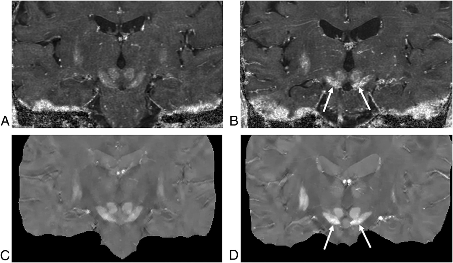
  • Fig 2.
    • Download figure
    • Open in new tab
    • Download powerpoint
    Fig 2.

    R2* (top row) and QSM maps (bottom row) of 2 subjects, a 68-year-old control subject (A and C) and a 66-year-old patient with PD (B and D). Both the R2* and the QSM maps show higher paramagnetic susceptibility in the substantia nigra of the patient with PD than in the control (arrows).
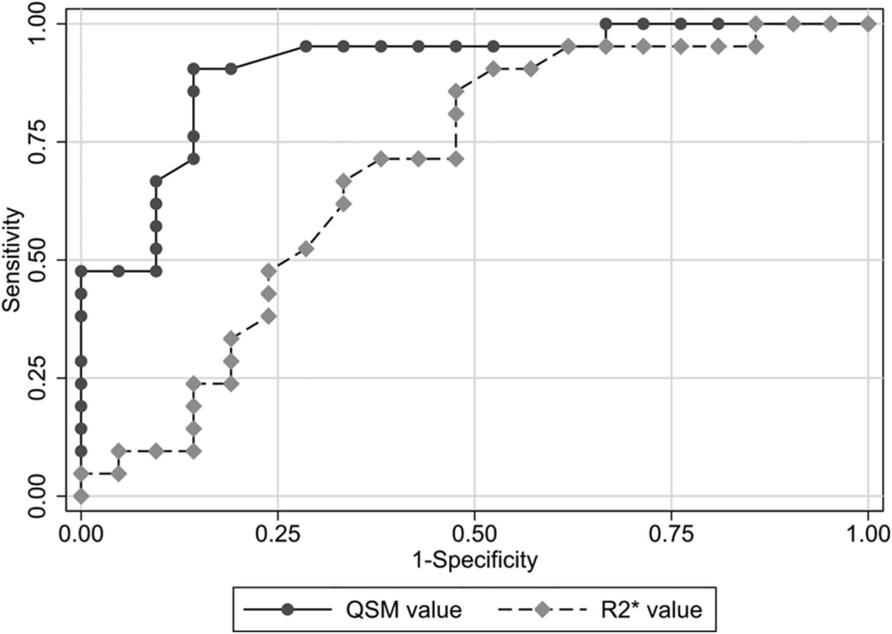
  • Fig 3.
    • Download figure
    • Open in new tab
    • Download powerpoint
    Fig 3.

    Graphs of the ROC curves for discriminating patients with PD and the controls. The Az value was 0.91 for QSMaverage, 0.90 for QSMmaximum, 0.69 for R2*average, and 0.71 for R2*maximum. Pair-wise comparisons showed that the Az for QSMaverage and QSMmaximum was significantly larger than that for R2*average and R2*maximum (P < .05).

Tables

  • Figures
    • View popup
    Table 1:

    Demographic and clinical data of subjects studied

    Patients with PD (n = 21)Controls (n = 21)P
    Sex (M/F)9:129:121.00
    Age (yr) (mean)72.0 ± 7.569.7 ± 8.6.54
    Onset (yr) (mean)69.0 ± 8.3
    Disease duration (mo) (mean)32.7 ± 27.1
    H & Y stage (median) (range)2 (1–3)
    • Note:—H & Y stage indicates Hoehn and Yahr stage.

    • View popup
    Table 2:

    Regional QSM and R2* values

    QSM (ppm)R2* Value (1/SE)
    Patients with PDControlsP ValuePatients with PDControlsP Value
    Substantia nigra0.224 ± 0.014a0.199 ± 0.014.01 × 10−330.1 ± 1.5b29.0 ± 2.0.01
    Red nucleus0.188 ± 0.0210.195 ± 0.019.1527.0 ± 1.727.8 ± 2.2.08
    Globus pallidus0.200 ± 0.0140.205 ± 0.029.8229.7 ± 1.829.5 ± 2.1.36
    Head of caudate0.166 ± 0.0130.166 ± 0.013.9725.2 ± 1.424.6 ± 1.7.19
    Putamen0.168 ± 0.0130.170 ± 0.009.1726.4 ± 0.926.7 ± 1.6.44
    Thalamus0.127 ± 0.0590.126 ± 0.007.5322.4 ± 1.022.8 ± 1.2.25
    • Note:—SE indicates standard error.

    • ↵a Significantly different from controls (P < .01).

    • ↵b Significantly different from controls (P < .05).

    • View popup
    Table 3:

    Mean number of sections and mean volume for the ROI measurements

    Mean No. of SectionsMean Volume (mm3)
    Patients with PDControlsPatients with PDControlsP Value
    Substantia nigra9.69.7598.2 ± 88.0612.5 ± 66.3.59
    Red nucleus4.84.7187.9 ± 15.3189.3 ± 16.6.87
    Globus pallidus19.019.12734.7 ± 263.02667.1 ± 241.6.54
    Head of caudate14.914.81529.9 ± 115.71564.6 ± 121.5.47
    Putamen18.418.44964.5 ± 505.25127.1 ± 718.4.62
    Thalamus15.215.11770.4 ± 89.71750.5 ± 118.4.63
    • View popup
    Table 4:

    Sensitivity, specificity, accuracy, and Az value of analyses of data in patients with PD and control subjectsa

    QSM AverageR2* Average
    Sensitivity90% (19/21)81% (17/21)
    Specificity86% (18/21)52% (11/21)
    Accuracy88% (37/42)67% (28/42)
    Az value0.91b0.69
    • ↵a Numbers in parentheses are raw data.

    • ↵b Significantly different from R2* (P < .05).

PreviousNext
Back to top

In this issue

American Journal of Neuroradiology: 36 (6)
American Journal of Neuroradiology
Vol. 36, Issue 6
1 Jun 2015
  • Table of Contents
  • Index by author
  • Complete Issue (PDF)
Advertisement
Print
Download PDF
Email Article

Thank you for your interest in spreading the word on American Journal of Neuroradiology.

NOTE: We only request your email address so that the person you are recommending the page to knows that you wanted them to see it, and that it is not junk mail. We do not capture any email address.

Enter multiple addresses on separate lines or separate them with commas.
Usefulness of Quantitative Susceptibility Mapping for the Diagnosis of Parkinson Disease
(Your Name) has sent you a message from American Journal of Neuroradiology
(Your Name) thought you would like to see the American Journal of Neuroradiology web site.
CAPTCHA
This question is for testing whether or not you are a human visitor and to prevent automated spam submissions.
Cite this article
Y. Murakami, S. Kakeda, K. Watanabe, I. Ueda, A. Ogasawara, J. Moriya, S. Ide, K. Futatsuya, T. Sato, K. Okada, T. Uozumi, S. Tsuji, T. Liu, Y. Wang, Y. Korogi
Usefulness of Quantitative Susceptibility Mapping for the Diagnosis of Parkinson Disease
American Journal of Neuroradiology Jun 2015, 36 (6) 1102-1108; DOI: 10.3174/ajnr.A4260

Citation Manager Formats

  • BibTeX
  • Bookends
  • EasyBib
  • EndNote (tagged)
  • EndNote 8 (xml)
  • Medlars
  • Mendeley
  • Papers
  • RefWorks Tagged
  • Ref Manager
  • RIS
  • Zotero
0 Responses
Respond to this article
Share
Bookmark this article
Usefulness of Quantitative Susceptibility Mapping for the Diagnosis of Parkinson Disease
Y. Murakami, S. Kakeda, K. Watanabe, I. Ueda, A. Ogasawara, J. Moriya, S. Ide, K. Futatsuya, T. Sato, K. Okada, T. Uozumi, S. Tsuji, T. Liu, Y. Wang, Y. Korogi
American Journal of Neuroradiology Jun 2015, 36 (6) 1102-1108; DOI: 10.3174/ajnr.A4260
del.icio.us logo Twitter logo Facebook logo Mendeley logo
  • Tweet Widget
  • Facebook Like
  • Google Plus One
Purchase

Jump to section

  • Article
    • Abstract
    • ABBREVIATIONS:
    • Materials and Methods
    • Results
    • Discussion
    • CONCLUSIONS
    • Footnotes
    • References
  • Figures & Data
  • Supplemental
  • Info & Metrics
  • Responses
  • References
  • PDF

Related Articles

  • PubMed
  • Google Scholar

Cited By...

  • Magnetic susceptibility is predictive of future clinical severity in Parkinsons disease
  • DeepQSM - Using Deep Learning to Solve the Dipole Inversion for MRI Susceptibility Mapping
  • Lateral Asymmetry and Spatial Difference of Iron Deposition in the Substantia Nigra of Patients with Parkinson Disease Measured with Quantitative Susceptibility Mapping
  • Crossref
  • Google Scholar

This article has not yet been cited by articles in journals that are participating in Crossref Cited-by Linking.

More in this TOC Section

  • Optimal MRI Sequence for Identifying Occlusion Location in Acute Stroke: Which Value of Time-Resolved Contrast-Enhanced MRA?
  • Evaluating the Effects of White Matter Multiple Sclerosis Lesions on the Volume Estimation of 6 Brain Tissue Segmentation Methods
  • Quiet PROPELLER MRI Techniques Match the Quality of Conventional PROPELLER Brain Imaging Techniques
Show more Brain

Similar Articles

Advertisement

Indexed Content

  • Current Issue
  • Accepted Manuscripts
  • Article Preview
  • Past Issues
  • Editorials
  • Editor's Choice
  • Fellows' Journal Club
  • Letters to the Editor
  • Video Articles

Cases

  • Case Collection
  • Archive - Case of the Week
  • Archive - Case of the Month
  • Archive - Classic Case

More from AJNR

  • Trainee Corner
  • Imaging Protocols
  • MRI Safety Corner
  • Book Reviews

Multimedia

  • AJNR Podcasts
  • AJNR Scantastics

Resources

  • Turnaround Time
  • Submit a Manuscript
  • Submit a Video Article
  • Submit an eLetter to the Editor/Response
  • Manuscript Submission Guidelines
  • Statistical Tips
  • Fast Publishing of Accepted Manuscripts
  • Graphical Abstract Preparation
  • Imaging Protocol Submission
  • Evidence-Based Medicine Level Guide
  • Publishing Checklists
  • Author Policies
  • Become a Reviewer/Academy of Reviewers
  • News and Updates

About Us

  • About AJNR
  • Editorial Board
  • Editorial Board Alumni
  • Alerts
  • Permissions
  • Not an AJNR Subscriber? Join Now
  • Advertise with Us
  • Librarian Resources
  • Feedback
  • Terms and Conditions
  • AJNR Editorial Board Alumni

American Society of Neuroradiology

  • Not an ASNR Member? Join Now

© 2025 by the American Society of Neuroradiology All rights, including for text and data mining, AI training, and similar technologies, are reserved.
Print ISSN: 0195-6108 Online ISSN: 1936-959X

Powered by HighWire