
Article Figures & Data
Figures
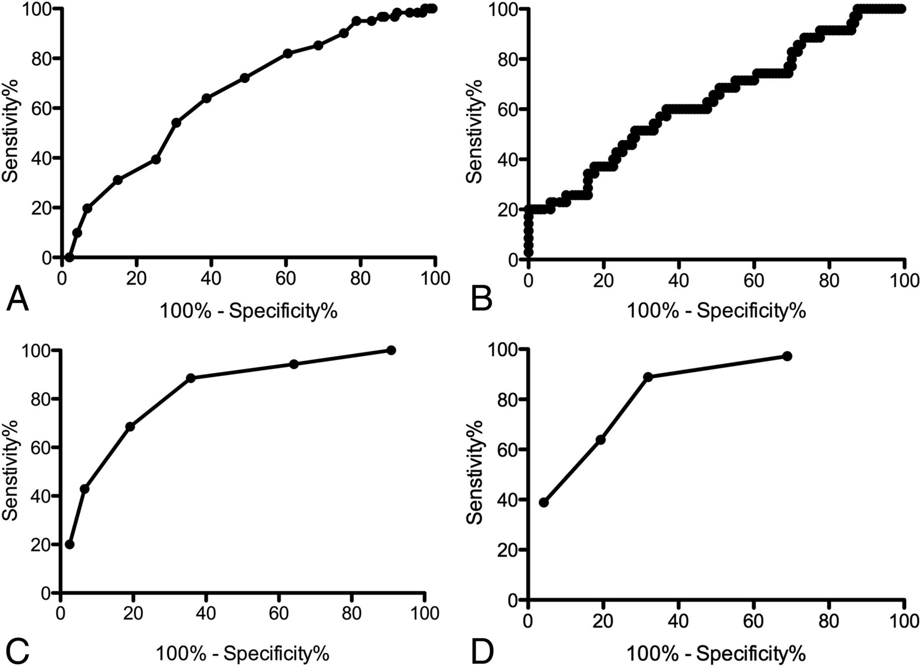
- Fig 1.
Receiver operating characteristic curves showing performance in predicting MGD for the largest lesion size (A), Wisconsin Index (B), the composite MGD score (C), and the 4D-CT MGD score (D).
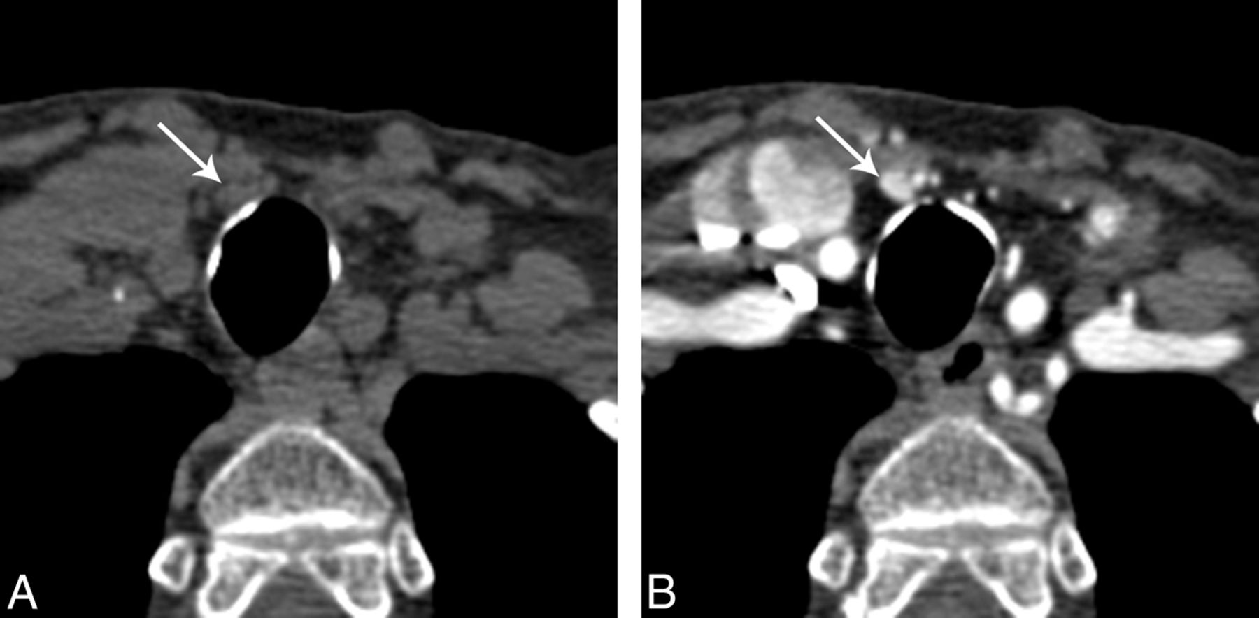
- Fig 2.
A 78-year-old woman with multigland disease, with a single small candidate lesion. A, Axial noncontrast CT just inferior to the thyroid gland shows a 6-mm nodule just deep to the strap muscles on the right (arrow). B, Axial arterial phase CT scan shows intense enhancement of this nodule. The serum calcium level was 10.1, and the serum parathyroid hormone level was 76. Despite identification of only a single lesion with 4D-CT, the composite MGD score was 4, and the probability of multigland disease was moderate. Surgical exploration revealed hyperplasia of the gland seen here and also hyperplastic bilateral superior parathyroid glands, neither of which could be seen even in retrospect.
- Fig 3.
A 67-year-old woman with single-gland disease with multiple prospective candidate lesions. A, Coronal arterial phase CT image shows an intensely enhancing nodule in an orthotopic left inferior gland location (long arrow), measuring 20 mm in diameter. B, Coronal arterial phase CT image posterior to A, viewed in the same window width/level, shows ovoid nodular lesions in the orthotopic right and left superior positions (short arrows). Despite appropriate location and shape, these lesions show less intense enhancement than is typical of parathyroid adenoma. The serum calcium level is 11.7, and the serum parathyroid hormone level is 211. Despite the presence of multiple candidate lesions, the composite MGD score is only 2 and the findings are predictive of single-gland disease.


Tables
Criterion Scoring No. of candidate lesions identified on 4D-CT Single lesion: 0 Multiple lesions: 2 No lesions: 2 Maximum diameter of largest lesion on 4D-CT >13 mm: 0 7–13 mm: 1 <7 mm or no lesion identified: 2 WIN >1600: 0 800–1600: 1 <800: 2 Note:—WIN indicates serum calcium level (milligram/deciliter) × serum parathyroid hormone level (picogram/milliliter).
↵a The composite MGD score includes all 3 components in the Table and ranges from 0 to 6. The 4D-CT MGD score does not include the Wisconsin Index and ranges from 0 to 4.
All subjects MGD SGD P Value No. of patients 155 36 119 No. of glands 216 97 119 Mean age (yr) 60 (range, 14–88) 59 60 Female 108 (67%) 25 (69%) 83 (70%) 4D-CT characteristics Mean size of abnormal glands (mm) 10.9 (5.9) 8.8 (4.0) 11.7 (6.4) .002 Median size of abnormal glands (mm) (IQR) 10 (7–13) 8 (6–11) 11 (7–13) No. <10 mm (%) 79 (37%) 39 (64%) 40 (34%) No. <7 mm (%) 30 (14%) 19 (31%) 11 (9%) No. >13 mm (%) 39 (18%) 6 (10%) 33 (28%) Prospectively detected lesions 1 100 10b 90 <.001 ≥2 46 20 26 None 9 6 3 Biochemical markers Serum calcium level (mg/dL) 11.0 (0.7) 10.8 (0.4) 11.1 (0.7) .07 Serum parathyroid hormone level (pg/mL) 117 (69) 92 (44) 122 (73) .02 WIN 1279 (744) 1005 (501) 1357 (783) .01 MGD scores Composite MGD score 2.6 (1.6) 4.1 (1.4) 2.2 (1.4) <.001 4D-CT MGD score 1.6 (1.4) 2.9 (1.1) 1.2 (1.2) <.001 4D-CT sensitivity based on original radiology reports Detection of lesions 167 (77%) 53 (55%) 114 (95%) Detection of all lesions in individual patients 126 (81%) 12 (33%) 114 (95%) - Table 3:
Performance of the composite MGD score for predicting MGD on the basis of the size of the largest lesion, the number of lesions prospectively identified, and the Wisconsin Indexa
MGD Score No. of Patients Sensitivity Specificity Positive Predictive Value ≥1 35 100% 9% 24% ≥2 33 94% 36% 30% ≥3 31 89% 64% 43% ≥4 24 69% 81% 51% ≥5 15 43% 93% 65% 6 7 20% 98% 70% ↵a There were no patients with scores of zero. One patient did not have recent serologic data.
- Table 4:
Performance of the 4D-CT MGD score for predicting MGD, based on the size of largest lesion and the number of lesions prospectively identified
MGD Score No. of Patients Sensitivity Specificity Positive Predictive Value 0 36 100% 0% 23% ≥1 35 97% 31% 30% ≥2 32 89% 68% 46% ≥3 23 64% 81% 50% 4 14 39% 96% 74%


