
Article Figures & Data
Figures
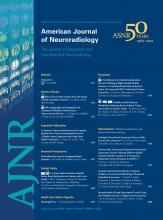
- Fig 1.
Tissue prolapse after stent implantation: an angiographic and 3D OCT evaluation. Angiography shows an inconclusive “haziness” inside the proximal edge of the stent (A, black arrow). B, 3D view of the stented carotid artery in which tissue prolapse is depicted (solid white arrows). The open-cell stent design (inlay with white background) is clearly demonstrated by the 3D image. The cross-section represented in C also shows tissue prolapse through the stent struts (solid white arrows). A good final angiographic result after the placement of a closed-cell stent is shown in D (black arrow). OCT 3D longitudinal reconstruction also reveals a different stent design (inlay with white background) and no significant remaining tissue prolapse (E), which can be confirmed by cross-sectional assessment (F).
- Fig 2.
TCFA rupture and inflammation visualized by OCT. A, Representative cross-sectional OCT image of a TCFA. The fibrous cap overlies a signal-intensity-poor region (white asterisks), corresponding to a lipid-rich plaque. B, The thickness (green double arrow) of the fibrous cap is 60 μm. A site of plaque rupture is shown in C, highlighting the presence of white thrombus (white arrow). D, The site of rupture also demonstrates bright spots that likely correspond to macrophage infiltration (dashed white circles) along the fibrous cap.
- Fig 3.
OCT 3D reconstruction of the right ICA. A, Moderate stenosis in the ICA is also demonstrated in the OCT longitudinal view in B. C and D, 3D OCT reconstructions reveal the region of interest along with proximal and distal reference segments. The numbered white dashed lines correspond to the cross-sectional images in E. A normal distal reference vessel is depicted in 1. The minimal luminal area (2) reveals a calcified plaque (solid white arrows). Signs of plaque instability are absent. The proximal reference area (3) exhibits a patent lumen with the presence of calcium (solid white arrows).
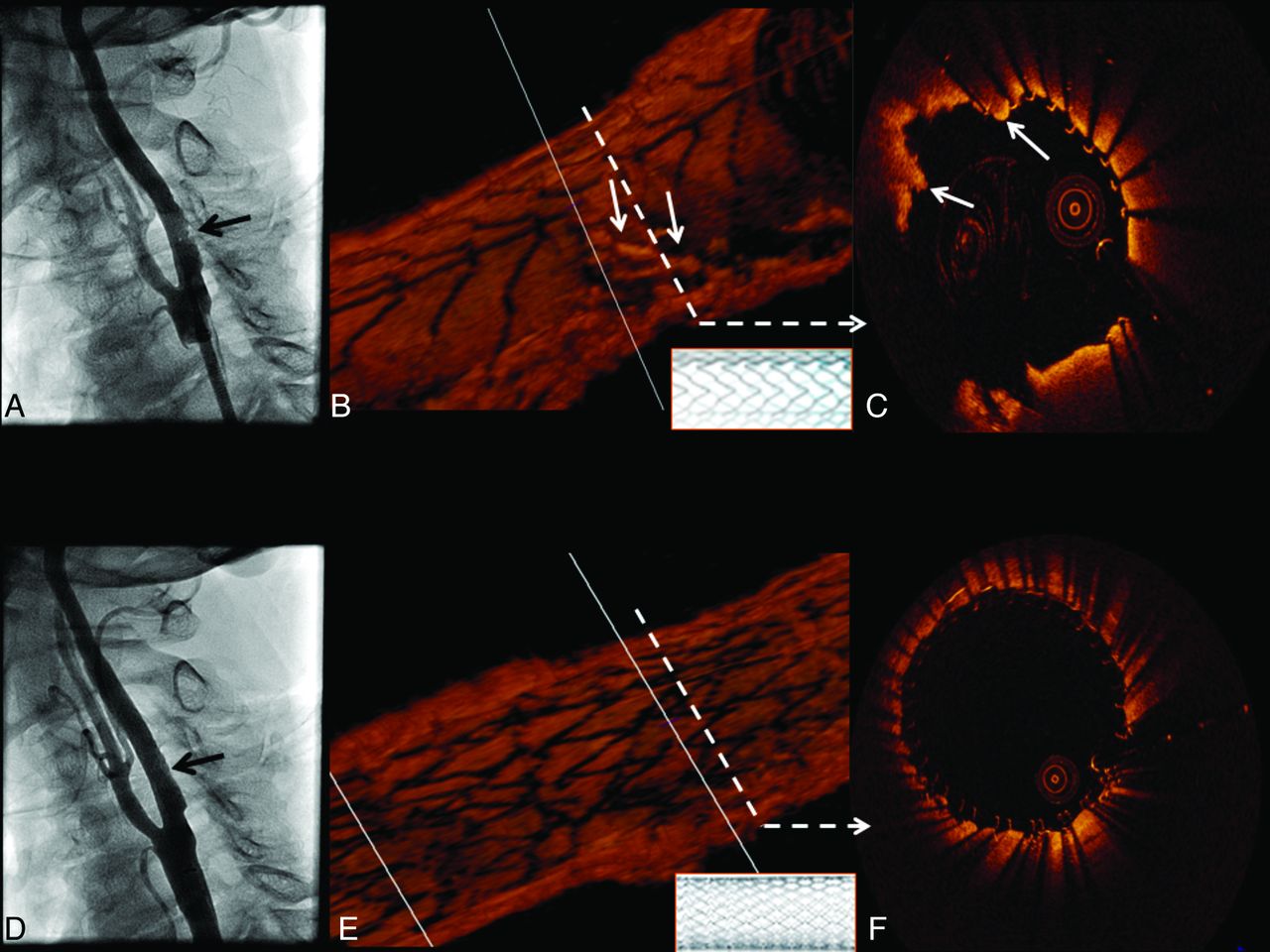
- Fig 4.
OCT evaluation of ICA in-stent restenosis. Angiography reveals ICA in-stent restenosis (A, black arrow), which is clearly depicted by longitudinal OCT assessment (B). The dashed white arrows correspond to the cross-sectional images in C, which reveal covered (white arrow) and uncovered (red arrow) stent struts. D, Heterogeneity of the pixel intensity in an area of neointimal hyperplasia (homogeneous high-intensity signal-intensity tissue, red arrows; and heterogeneous predominantly low-intensity signal-intensity tissue, white arrows). Furthermore, intraluminal thrombus is revealed (D, blue arrow). After stent placement, angiography shows a good result (E). OCT cross-sectional image (F) depicts 2 layers of stent struts: the previously deployed (blue dashed arrow) and the recently implanted stent (blue solid arrow).
- Fig 5.
OCT demonstration of ICA calcified plaque, TCFA, and plaque ulceration. A, Carotid angiogram shows a mild-to-moderate narrowing of the right ICA (region of interest, black dashed square). OCT longitudinal view of the region of interest is revealed in B. The white dashed arrows point to their respective cross-sectional images. C, Highly calcified plaque is shown (white asterisks). The same cross-section is represented in D, using semiautomated software for calcium detection and quantification (highlighted in yellow). E, TCFA. A signal-intensity-poor region (white asterisks) corresponding to lipid presence is identified below the fibrous cap (white arrowheads). Automated fibrous cap thickness detection F, Fibrous cap heterogeneity in a color-coded display (pink, <65 μm; green, 65–149 μm; blue, 150–220 μm). G, Red dashed circle represents cavity formation due to plaque ulceration. This area is also easily identified in the longitudinal view (B). Another cavity (H, red asterisk) located more proximally is also noted.


In this issue


Jump to section
Related Articles
Cited By...
- Optical Coherence Tomography: Future Applications in Cerebrovascular Imaging
- Optical coherence tomography evaluation of tissue prolapse after carotid artery stenting using closed cell design stents for unstable plaque
- Optical coherence tomography of the intracranial vasculature and Wingspan stent in a patient
- Intravascular Frequency-Domain Optical Coherence Tomography Assessment of Carotid Artery Disease in Symptomatic and Asymptomatic Patients
- Optical coherence tomography of the intracranial vasculature and Wingspan stent in a patient
- Frequency-Domain Optical Coherence Tomography Assessment of Human Carotid Atherosclerosis Using Saline Flush for Blood Clearance without Balloon Occlusion