Advertisement

Main menu

  • Home
  • Content
    • Current Issue
    • Accepted Manuscripts
    • Article Preview
    • Past Issue Archive
    • Video Articles
    • AJNR Case Collection
    • Case of the Week Archive
    • Case of the Month Archive
    • Classic Case Archive
  • Special Collections
    • AJNR Awards
    • Low-Field MRI
    • Alzheimer Disease
    • ASNR Foundation Special Collection
    • Photon-Counting CT
    • View All
  • Multimedia
    • AJNR Podcasts
    • AJNR SCANtastic
    • Trainee Corner
    • MRI Safety Corner
    • Imaging Protocols
  • For Authors
    • Submit a Manuscript
    • Submit a Video Article
    • Submit an eLetter to the Editor/Response
    • Manuscript Submission Guidelines
    • Statistical Tips
    • Fast Publishing of Accepted Manuscripts
    • Graphical Abstract Preparation
    • Imaging Protocol Submission
    • Author Policies
  • About Us
    • About AJNR
    • Editorial Board
    • Editorial Board Alumni
  • More
    • Become a Reviewer/Academy of Reviewers
    • Subscribers
    • Permissions
    • Alerts
    • Feedback
    • Advertisers
    • ASNR Home

User menu

  • Alerts
  • Log in

Search

  • Advanced search
American Journal of Neuroradiology

American Journal of Neuroradiology

ASHNR American Society of Functional Neuroradiology ASHNR American Society of Pediatric Neuroradiology ASSR
  • Alerts
  • Log in

Advanced Search

  • Home
  • Content
    • Current Issue
    • Accepted Manuscripts
    • Article Preview
    • Past Issue Archive
    • Video Articles
    • AJNR Case Collection
    • Case of the Week Archive
    • Case of the Month Archive
    • Classic Case Archive
  • Special Collections
    • AJNR Awards
    • Low-Field MRI
    • Alzheimer Disease
    • ASNR Foundation Special Collection
    • Photon-Counting CT
    • View All
  • Multimedia
    • AJNR Podcasts
    • AJNR SCANtastic
    • Trainee Corner
    • MRI Safety Corner
    • Imaging Protocols
  • For Authors
    • Submit a Manuscript
    • Submit a Video Article
    • Submit an eLetter to the Editor/Response
    • Manuscript Submission Guidelines
    • Statistical Tips
    • Fast Publishing of Accepted Manuscripts
    • Graphical Abstract Preparation
    • Imaging Protocol Submission
    • Author Policies
  • About Us
    • About AJNR
    • Editorial Board
    • Editorial Board Alumni
  • More
    • Become a Reviewer/Academy of Reviewers
    • Subscribers
    • Permissions
    • Alerts
    • Feedback
    • Advertisers
    • ASNR Home
  • Follow AJNR on Twitter
  • Visit AJNR on Facebook
  • Follow AJNR on Instagram
  • Join AJNR on LinkedIn
  • RSS Feeds

AJNR Awards, New Junior Editors, and more. Read the latest AJNR updates

  • Getting new auth cookie, if you see this message a lot, tell someone!
  • Getting new auth cookie, if you see this message a lot, tell someone!
Review ArticleReview Articles
Open Access

Syndromes of the First and Second Branchial Arches, Part 2: Syndromes

J.M. Johnson, G. Moonis, G.E. Green, R. Carmody and H.N. Burbank
American Journal of Neuroradiology February 2011, 32 (2) 230-237; DOI: https://doi.org/10.3174/ajnr.A2073
J.M. Johnson
  • Find this author on Google Scholar
  • Find this author on PubMed
  • Search for this author on this site
G. Moonis
  • Find this author on Google Scholar
  • Find this author on PubMed
  • Search for this author on this site
G.E. Green
  • Find this author on Google Scholar
  • Find this author on PubMed
  • Search for this author on this site
R. Carmody
  • Find this author on Google Scholar
  • Find this author on PubMed
  • Search for this author on this site
H.N. Burbank
  • Find this author on Google Scholar
  • Find this author on PubMed
  • Search for this author on this site
  • Article
  • Figures & Data
  • Info & Metrics
  • Responses
  • References
  • PDF
Loading

Article Figures & Data

Figures

  • Fig 1.
    • Download figure
    • Open in new tab
    • Download powerpoint
    Fig 1.

    Young adult with hemifacial microstomia. A−C, 3D bone reconstruction shows right mandibular and maxillary hypoplasia compared with the normal-appearing left condyle. D, A 3-year-old boy with hemifacial microstomia. 3D bone reconstruction shows a more dramatic appearance of asymmetric hypoplasia of the mandible.
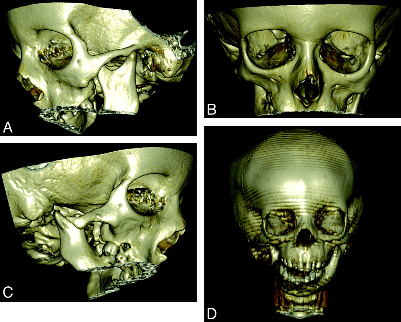
  • Fig 2.
    • Download figure
    • Open in new tab
    • Download powerpoint
    Fig 2.

    A 5-year-old boy with ACS. A−C, 3D bony reconstructions show an absent condylar processes, asymmetric micrognathia, and hypoplastic condyles. D, Axial CT scan shows bilateral abnormal TMJs (arrows) with dysplastic condylar processes.
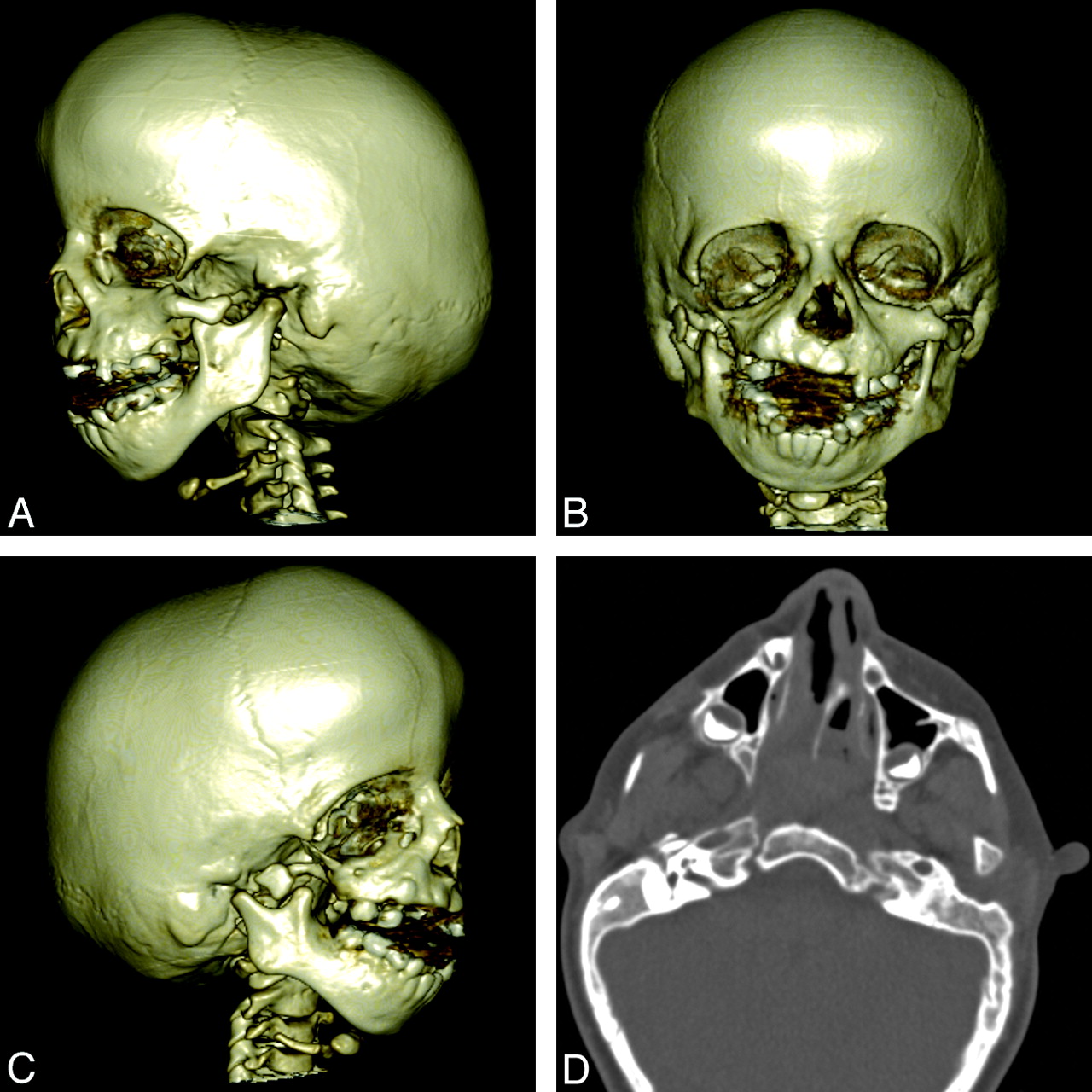
  • Fig 3.
    • Download figure
    • Open in new tab
    • Download powerpoint
    Fig 3.

    An 18-year-old man with ACS. A, 3D bony reconstruction shows micrognathia with overprotrusion of the mandible in relation to the maxilla. The lateral mandibular cleft is a feature commonly seen in ACS (see also Fig 2). B, Axial CT scan also shows the asymmetry of the dysplastic protruding mandible.
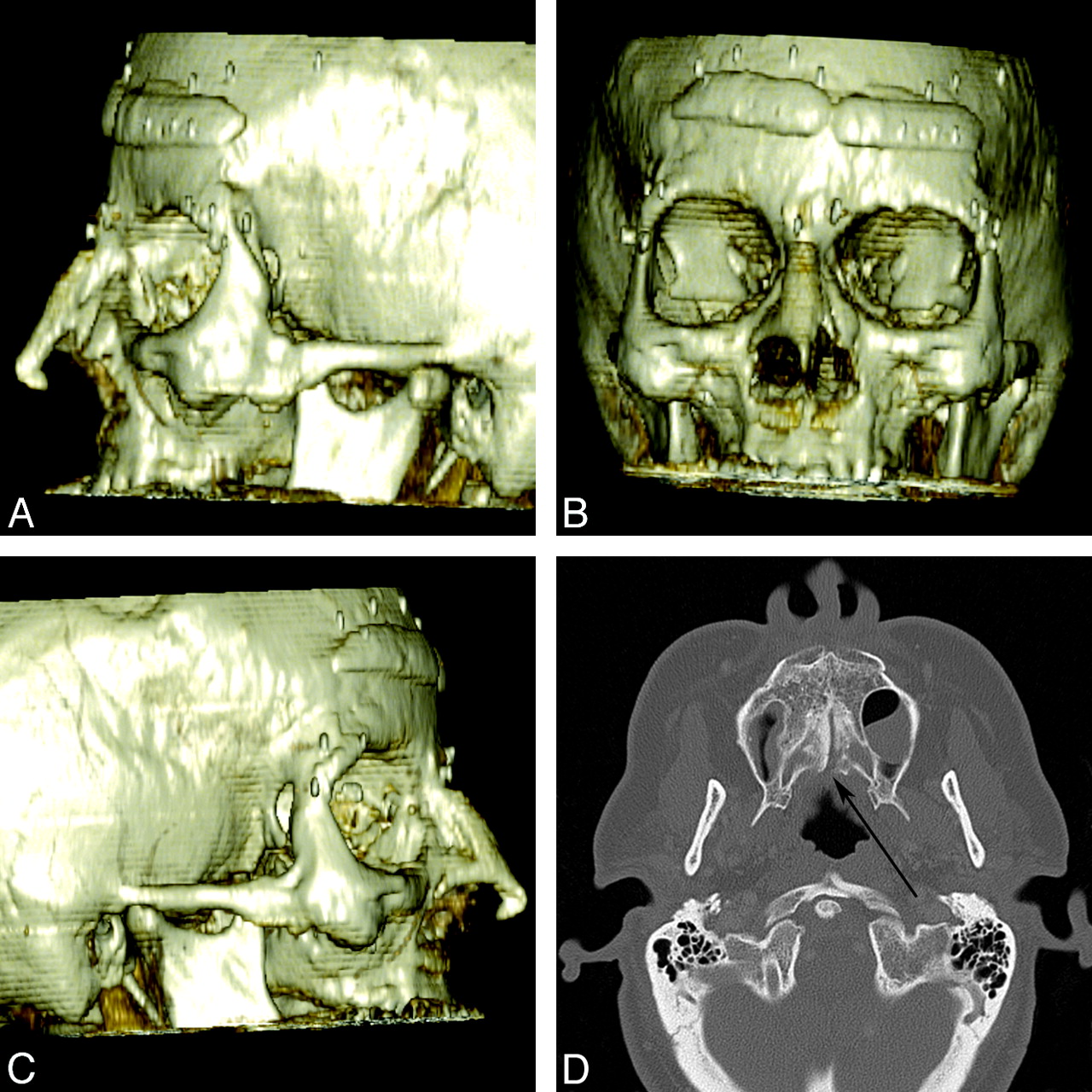
  • Fig 4.
    • Download figure
    • Open in new tab
    • Download powerpoint
    Fig 4.

    A 4-year-old girl with TCS. A−C, 3D bone reconstructions show bilateral and asymmetric abnormalities of the mandibular condyle and coronoid process as well as maxillary hypoplasia and micrognathia. The EACs are absent bilaterally. D, Axial CT image shows bilateral hypoplastic zygomatic arches, maxillary bone dysplasia, and temporal bone abnormalities.

  • Fig 5.
    • Download figure
    • Open in new tab
    • Download powerpoint
    Fig 5.

    A 47-year-old woman with Stickler syndrome. A−C, 3D bony reconstructions show a flat midface with a depressed nasal bridge, short nose, anteverted nares, and zygomatic hypoplasia. Reconstructive and cosmetic hardware and implants are present. D, Axial CT scan shows incomplete fusion of the palatal bones with a posterior submucosal cleft (arrow).

  • Fig 6.
    • Download figure
    • Open in new tab
    • Download powerpoint
    Fig 6.

    A 6-year-old boy with Stickler syndrome. 3D bony reconstructions show the characteristic flat midface with a depressed nasal bridge. The degree of zygomatic hypoplasia is more subtle. There is mild micrognathia. An endotracheal tube is present. Courtesy of Michael Cunningham.

  • Fig 7.
    • Download figure
    • Open in new tab
    • Download powerpoint
    Fig 7.

    A 6-year-old boy with VCFS. A−C, 3D bone reconstructions show mild micrognathia with normal condyle and coronoid process morphology. D, Axial CT angiogram shows relatively symmetric micrognathia and areas of malocclusion (arrows).

PreviousNext
Back to top

In this issue

American Journal of Neuroradiology: 32 (2)
American Journal of Neuroradiology
Vol. 32, Issue 2
1 Feb 2011
  • Table of Contents
  • Index by author
Advertisement
Print
Download PDF
Email Article

Thank you for your interest in spreading the word on American Journal of Neuroradiology.

NOTE: We only request your email address so that the person you are recommending the page to knows that you wanted them to see it, and that it is not junk mail. We do not capture any email address.

Enter multiple addresses on separate lines or separate them with commas.
Syndromes of the First and Second Branchial Arches, Part 2: Syndromes
(Your Name) has sent you a message from American Journal of Neuroradiology
(Your Name) thought you would like to see the American Journal of Neuroradiology web site.
CAPTCHA
This question is for testing whether or not you are a human visitor and to prevent automated spam submissions.
Cite this article
J.M. Johnson, G. Moonis, G.E. Green, R. Carmody, H.N. Burbank
Syndromes of the First and Second Branchial Arches, Part 2: Syndromes
American Journal of Neuroradiology Feb 2011, 32 (2) 230-237; DOI: 10.3174/ajnr.A2073

Citation Manager Formats

  • BibTeX
  • Bookends
  • EasyBib
  • EndNote (tagged)
  • EndNote 8 (xml)
  • Medlars
  • Mendeley
  • Papers
  • RefWorks Tagged
  • Ref Manager
  • RIS
  • Zotero
0 Responses
Respond to this article
Share
Bookmark this article
Syndromes of the First and Second Branchial Arches, Part 2: Syndromes
J.M. Johnson, G. Moonis, G.E. Green, R. Carmody, H.N. Burbank
American Journal of Neuroradiology Feb 2011, 32 (2) 230-237; DOI: 10.3174/ajnr.A2073
del.icio.us logo Twitter logo Facebook logo Mendeley logo
  • Tweet Widget
  • Facebook Like
  • Google Plus One
Purchase

Jump to section

  • Article
    • Abstract
    • Abbreviations
    • Syndromes
    • Conclusions
    • Acknowledgments
    • References
  • Figures & Data
  • Info & Metrics
  • Responses
  • References
  • PDF

Related Articles

  • No related articles found.
  • PubMed
  • Google Scholar

Cited By...

  • No citing articles found.
  • Crossref
  • Google Scholar

This article has not yet been cited by articles in journals that are participating in Crossref Cited-by Linking.

More in this TOC Section

  • An Atlas of Neonatal Neurovascular Imaging Anatomy as Depicted with Microvascular Imaging: The Intracranial Arteries
  • An Atlas of Neonatal Neurovascular Imaging Anatomy as Depicted with Microvascular Imaging: The Intracranial Veins
  • Clinical Translation of Hyperpolarized 13C Metabolic Probes for Glioma Imaging
Show more Review Articles

Similar Articles

Advertisement

Indexed Content

  • Current Issue
  • Accepted Manuscripts
  • Article Preview
  • Past Issues
  • Editorials
  • Editor's Choice
  • Fellows' Journal Club
  • Letters to the Editor
  • Video Articles

Cases

  • Case Collection
  • Archive - Case of the Week
  • Archive - Case of the Month
  • Archive - Classic Case

More from AJNR

  • Trainee Corner
  • Imaging Protocols
  • MRI Safety Corner
  • Book Reviews

Multimedia

  • AJNR Podcasts
  • AJNR Scantastics

Resources

  • Turnaround Time
  • Submit a Manuscript
  • Submit a Video Article
  • Submit an eLetter to the Editor/Response
  • Manuscript Submission Guidelines
  • Statistical Tips
  • Fast Publishing of Accepted Manuscripts
  • Graphical Abstract Preparation
  • Imaging Protocol Submission
  • Evidence-Based Medicine Level Guide
  • Publishing Checklists
  • Author Policies
  • Become a Reviewer/Academy of Reviewers
  • News and Updates

About Us

  • About AJNR
  • Editorial Board
  • Editorial Board Alumni
  • Alerts
  • Permissions
  • Not an AJNR Subscriber? Join Now
  • Advertise with Us
  • Librarian Resources
  • Feedback
  • Terms and Conditions
  • AJNR Editorial Board Alumni

American Society of Neuroradiology

  • Not an ASNR Member? Join Now

© 2025 by the American Society of Neuroradiology All rights, including for text and data mining, AI training, and similar technologies, are reserved.
Print ISSN: 0195-6108 Online ISSN: 1936-959X

Powered by HighWire