
Article Figures & Data
Figures
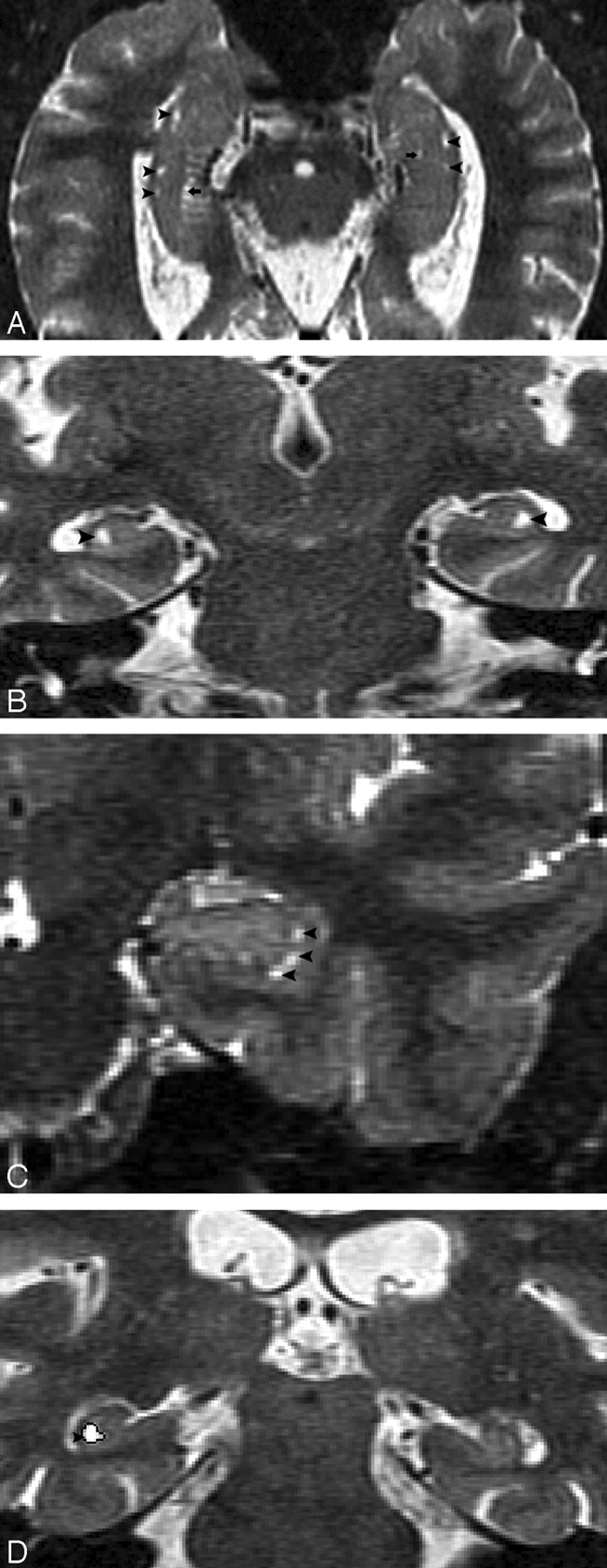
- Fig 1.
MR imaging appearance of hippocampal sulcus residual cavity (HSC).
A, The image was reformatted to a 1-mm-thick axial plane from the coronal T2 short-τ inversion recovery (STIR) scan, paralleling to the long axial of hippocampus (HSC, arrowheads; perihippocampal fissures [PHF], arrows).
B–D, Images were obtained by using the T2 STIR sequence (section thickness, 3 mm; HSC, arrowheads).
D, Region of interest of HSC (arrowhead).
- Fig 2.
A and B, Normal anatomy of perihippocampal fissure.
A, Axial diagram showing the structure of perihippocampal fissures (PHF).
B, Coronal diagram showing the structure of PHF.
Black arrows, PHF; black arrowhead, hippocampal fissure; white arrowhead, choroidal fissure; curved arrow, transverse fissure of Bichat); H, hippocampus; S, subiculum; AC, ambient cistern.
- Fig 3.
Four-point subjective rating scale of the perihippocampal fissures (PHF).
A and B, Axial and coronal view of same case. Left = 0; right = 0
C and D, Axial and coronal view of same case. Left = 1; right = 1
E and F, Axial and coronal view of same case. Left = 2; right = 2
G and H, Axial and coronal view of same case. Left = 3; right = 3
Axial images were reformatted in 1-mm thickness from inferior to superior, paralleling to the long axial of hippocampus. Black arrows highlight the PHF. There are 3 coronal views for each case showing the head, bodyi and tail of hippocampus. Black arrows highlight the PHF. White arrows on coronal plane show lateral geniculate body. Arrowheads on coronal plane show uncal sulcus.
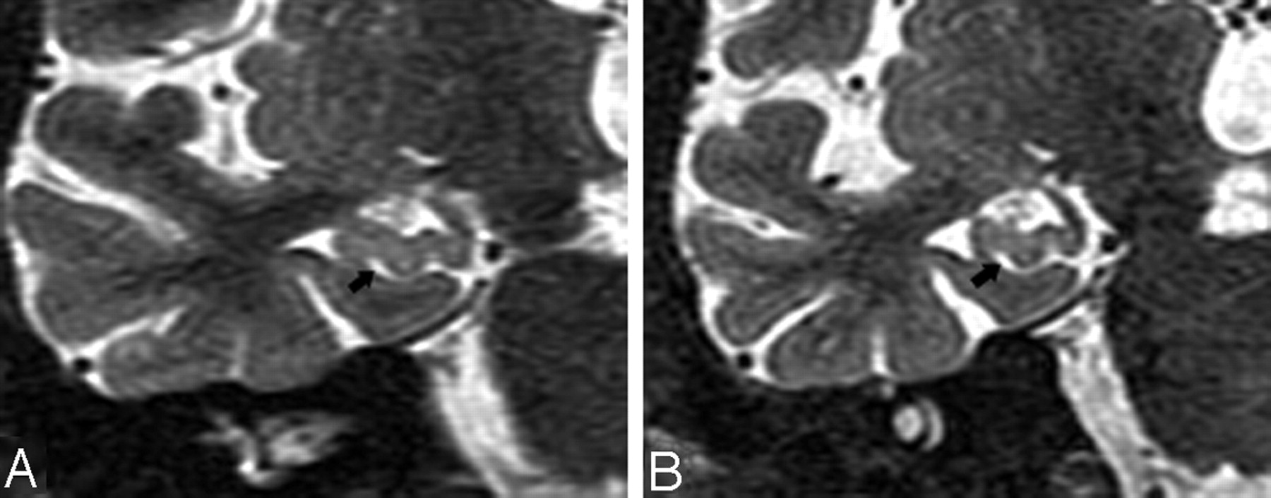
- Fig 4.
T2 short-τ inversion recovery (STIR) MR coronal image obtained from a 74-year-old male normal control (NC) patient. A, Arrowhead shows a small hippocampal sulcus residual cavity (HSC); arrow shows perihippocampal fissures (PHF) (uncal sulcus). B, 3-year follow-up scan. Comparing A and B, there is progressive dilation of the uncal sulcus (arrow), but no obvious change in the size of the HSC (arrowhead).
- Fig 5.
T2 short-τ inversion recovery (STIR) MR coronal image was obtained from a 73-year-old male normal control (NC) patient. Arrows show a dilated uncal sulcus and no hippocampal sulcus residual cavity (HSC). B, 3-year follow-up.
- Fig 6.
T2 short-τ inversion recovery (STIR) MR image was obtained from a 66-year-old female normal control (NC) patient.
A, Axial plane, arrowheads show the hippocampal sulcus residual cavity (HSC) and arrow shows the uncal sulcus. We can best differentiate them on the coronal plane.
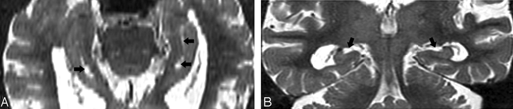
- Fig 7.
Image obtained from a 90-year-old female normal control (NC) patient. Axial (A) and coronal (B) T2 short-τ inversion recovery (STIR) MR image show the perihippocampal fissures (PHF) (black arrows). This is also best seen along the coronal plane (B).
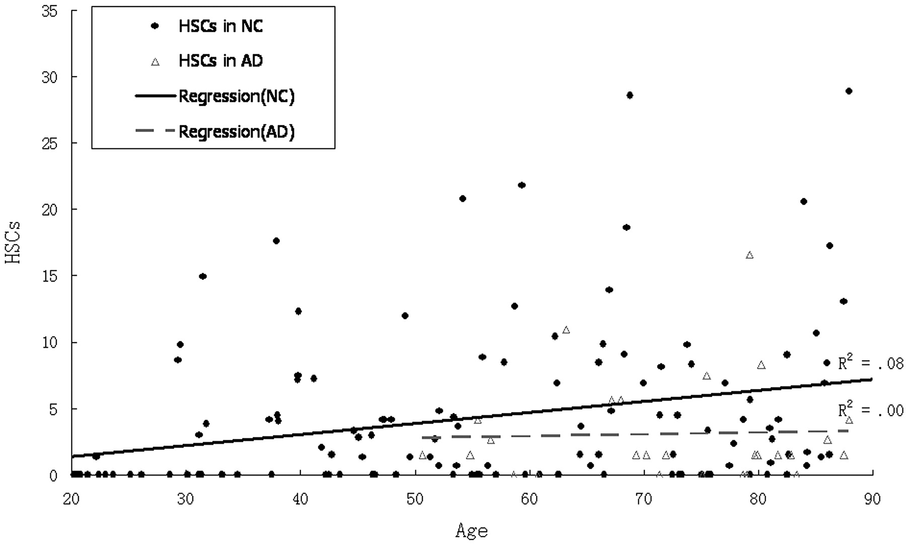
- Fig 8.
Scatter plot and regression lines show the relationship between age and hippocampal sulcus residual cavity (HSC) in both normal control (NC) patients and those with Alzheimer disease (AD).
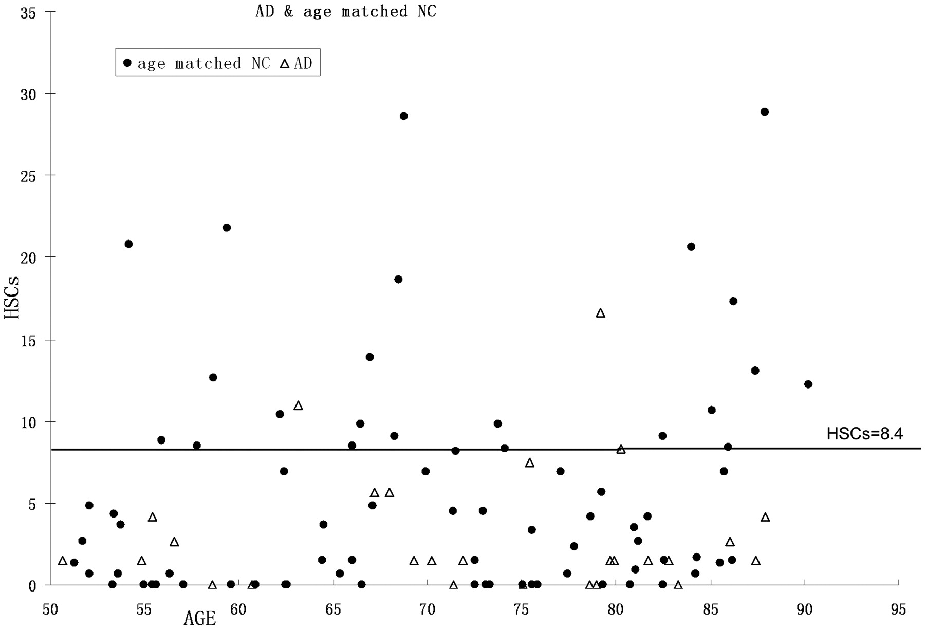
- Fig 9.
Scatter plot and cutoff (hippocampal sulcus residual cavity [HSC], 8.4) for the top 20% high HSC cases.
- Fig 10.
Scatter plot and regression lines showing the relationship between age and perihippocampal fissures (PHF) in both normal control (NC) patients and Alzheimer disease (AD).
- Fig 11.
Images derived from the human hippocampus (Duvernoy, 1988)5.
A, Six-week-old embryo. Open arrow shows the primitive hippocampal sulcus.
B, Adult. Black arrow shows the hippocampal fissure; arrowhead shows residual cavity.
C, T2 coronal MR image was obtained from a 67-year-old female normal control (NC) patient. Arrowhead shows the same shape hippocampal sulcus residual cavity (HSC) as in B.
- Fig 12.
T2 MR imaging (A) and the corresponding postmortem gross specimen (B) from an 86-year-old woman with Alzheimer disease (AD), showing a crescent-shaped HSC (arrowhead) and an enlarged uncal sulcus (arrow).


Tables
Age Group (y) N Female (%) Education (y; mean ± SD) Age (y; mean ± SD) 20–30 18 56 15 ± 1.6 24 ± 3.6 31–40 17 53 17 ± 1.9 35 ± 3.4 41–50 19 58 17 ± 1.5 46 ± 2.6 51–60 19 58 17 ± 1.4 55 ± 2.6 61–70 18 50 16 ± 2.3 66 ± 2.6 71–80 19 53 16 ± 2.5 75 ± 2.6 81–90 20 65 15 ± 2.2 84 ± 2.6 50–88* 27 67 16 ± 2.7 72 ± 10.8 Note:—Normal controls in age groups 20–90 = 130.
* Alzheimer disease group.
Age Group (y) N Cavity-Positive Finding Patients with HSC (%) Patients with Bilateral HSC (%) Patients with Multiple HSC (%) 20–30 18 22 17 17 31–40 17 59 47 47 41–50 19 63 32 37 51–60 19 68 37 42 61–70 18 78 56 56 71–80 19 63 21 58 81–90 20 90 55 55 Total 130 64 38 42 Note:—HSC indicates hippocampal salcus residual cavity.
N HSCs (mean ± SD) PHFs (mean ± SD) Normal controls 130 4.37 ± 5.97 1.53 ± 1.47 Alzheimer disease 27 3.08 ± 3.92 4.89 ± 1.22 Age-matched normal controls 62 5.54 ± 4.39 2.25 ± 1.48 Note:—HSC indicates hippocampal sulcus residual cavity; PHF, perihippocampal fissure.
Age-Matched Normal Controls Alzheimer Disease Total >8.4 mm2 21 2 23 <8.4 mm2 55 25 80 Total 76 27 103 Note:—HSC indicates hippocampal sulcus residual cavity.
Study Frequency of HSC (%) Age (y) Sequence Orientation Thickness (mm) Gap (mm) Matrix Sasaki et al6 39 8–85 T2 SE Axial 5–7 * 256 × 192 Yoneoka et al13 26 18–80 T2 FSE Coronal 5 2.5 512 × 512 Bartres-Faz et al12 93 50–85 T2 FSE Axial 3 * 256 × 256 Current study 64 20–90 T2 STIR Coronal 3 1 512 × 256 Note:—HSC indicates hippocampal sulcus residual cavity.
* No mention.
In this issue


Jump to section
Related Articles
Cited By...
- No citing articles found.