
Article Figures & Data
Figures
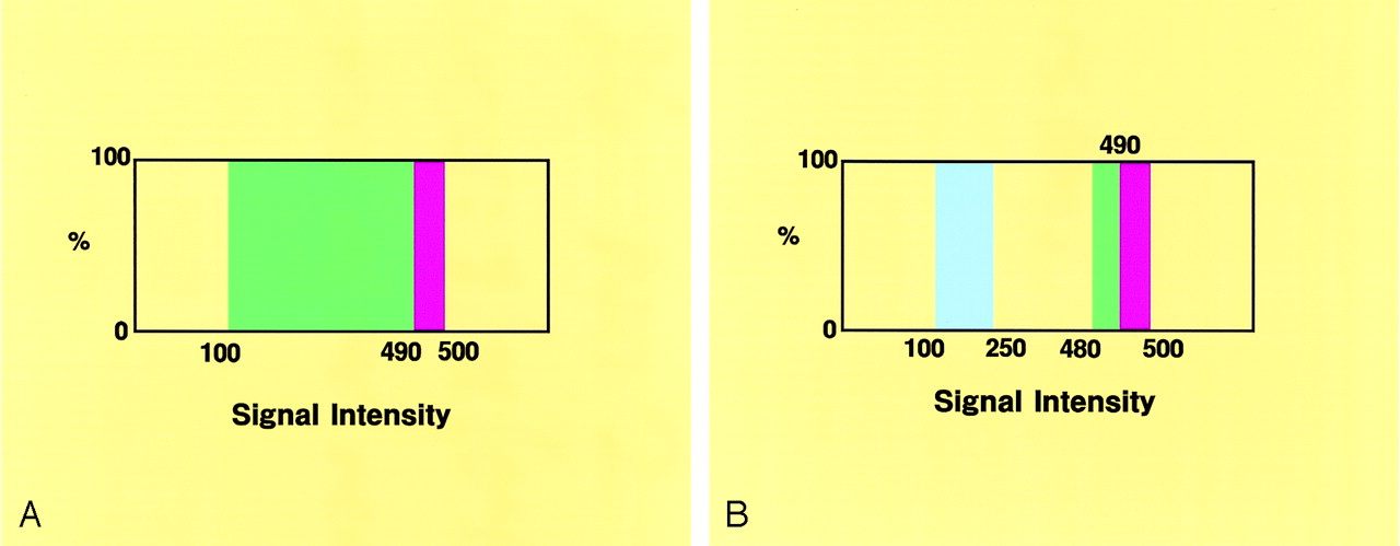
- Fig 1.
Opacity charts of the signal intensity distribution for the 3D MR cisternograms.
A, Opacity chart for the conventional 3D MR cisternogram, showing a square curve with a threshold range of 100–500 (100% opacity level), and color-rendered in green (threshold range 100–490) and red (490–500).
B, Opacity chart for the transparent 3D MR cisternogram, showing square curves, one with a threshold range of 480–500 (100% opacity level), color-rendered in green (threshold range 480–490) and red (490–500), and the other with a threshold range of 100–250 (100% opacity level), color-rendered in blue.
- Fig 2.
Case 1, an 83-year-old woman with a large unruptured right internal carotid–posterior communicating artery aneurysm and a small anterior choroidal artery aneurysm.
A, Operative photograph showing the caudal view of the right superolateral aspect of the posterior communicating artery aneurysm (An 1), the anterior choroidal artery aneurysm (An 2), and the right optic nerve (ON).
B, Digital subtraction angiogram, right oblique projection, showing the aneurysms (An 1, An 2).
C, 3D MR angiogram, similar projection to the operative view in panel A, showing the arterial components of the aneurysmal complex. Note the slightly concave surface at the neck (arrowheads).
D, MinIP image obtained from MR cisternography, superoinferior projection, showing the cisternal structures with negative shadows in contrast to the surrounding CSF with a positive shadow. ON, optic nerve; AC, anterior clinoid process; PC, posterior clinoid process; PF, petroclinoid dural fold.
E, Conventional 3D MR cisternogram, similar projection to the operative view in panel A, depicting the contours of the aneurysmal complex (An 1, An 2, A1, C2, M1), the right optic nerve (ON), the anterior clinoid process (AC), the petroclinoidal dural fold (PF), and the temporal lobe (TL).
F, Conventional 3D MR cisternogram, virtual viewpoint from superoposterior projection, showing the aneurysmal complex and perianeurysmal environment.
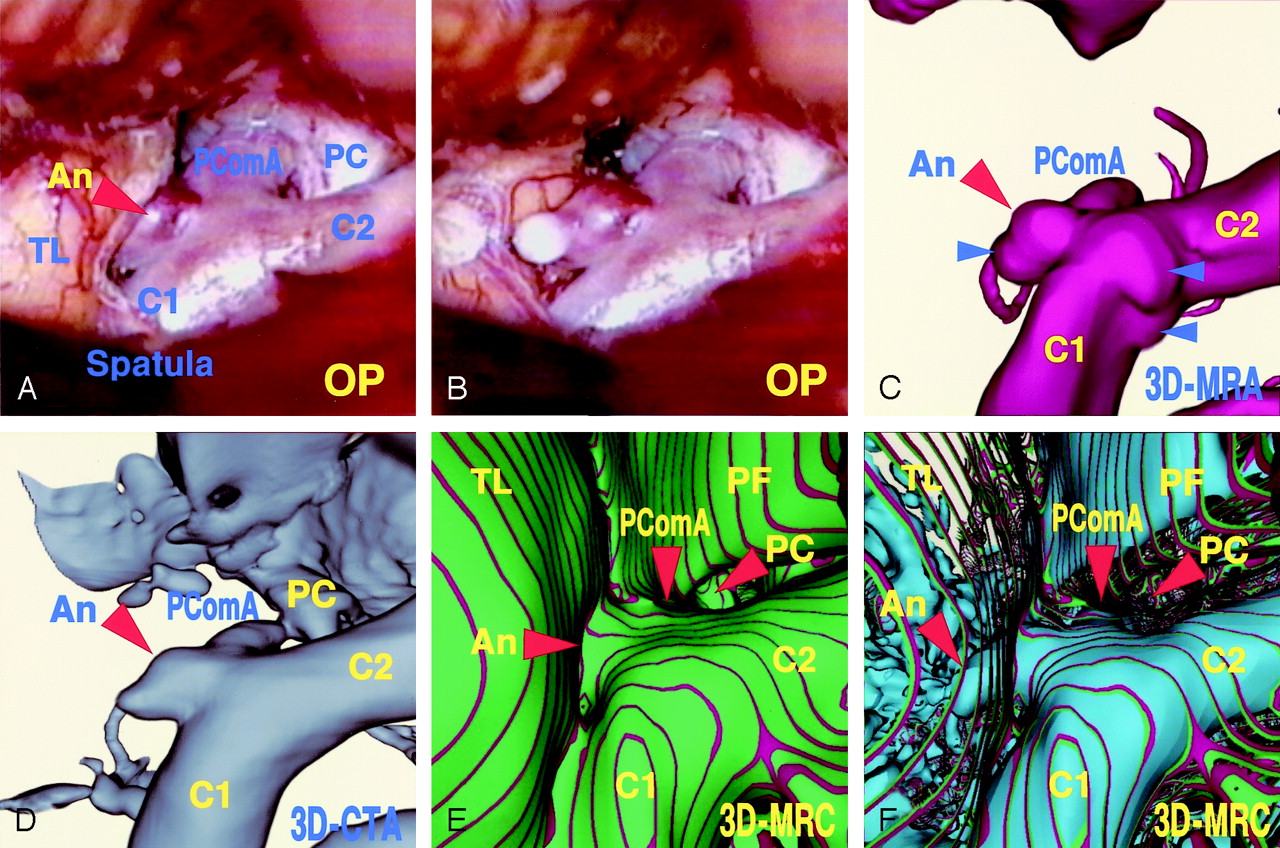
- Fig 3.
Case 2, a 54-year-old man with an unruptured left internal carotid–posterior communicating artery aneurysm.
Operative photographs before (A) and after (B) surgical dissection of the embedded aneurysmal dome, showing the aneurysm (An), the internal carotid artery (C1, C2), the posterior communicating artery (PComA), posterior clinoid process (PC), and the temporal lobe (TL).
C, 3D MR angiogram, similar projection to the operative views in panels A and B, showing the aneurysmal complex. Note the slight difference in terms of the aneurysmal shape and small protrusions on the parent artery (arrowheads).
D, 3D CT angiogram, similar projection with the operative views in panels A and B, showing the aneurysmal complex and cranial base bone similarly.
E, Conventional 3D MR cisternogram, similar projection to the operative views in panels A and B, depicting the contours of the aneurysmal complex (An, C1, C2, PComA), the posterior clinoid process bone (PC), the petroclinoidal dural fold (PF), and the temporal lobe (TL).
F, Transparent 3D MR cisternogram, the same projection as in panel E, depicting the whole shape of the aneurysm (An) and parent arteries (C1, C2, PComA) in blue, transparently through the vessel wall and the adjacent brain surface (green and red).


