Advertisement

Main menu

  • Home
  • Content
    • Current Issue
    • Accepted Manuscripts
    • Article Preview
    • Past Issue Archive
    • Video Articles
    • AJNR Case Collection
    • Case of the Week Archive
    • Case of the Month Archive
    • Classic Case Archive
  • Special Collections
    • AJNR Awards
    • Low-Field MRI
    • Alzheimer Disease
    • ASNR Foundation Special Collection
    • Photon-Counting CT
    • View All
  • Multimedia
    • AJNR Podcasts
    • AJNR SCANtastic
    • Trainee Corner
    • MRI Safety Corner
    • Imaging Protocols
  • For Authors
    • Submit a Manuscript
    • Submit a Video Article
    • Submit an eLetter to the Editor/Response
    • Manuscript Submission Guidelines
    • Statistical Tips
    • Fast Publishing of Accepted Manuscripts
    • Graphical Abstract Preparation
    • Imaging Protocol Submission
    • Author Policies
  • About Us
    • About AJNR
    • Editorial Board
    • Editorial Board Alumni
  • More
    • Become a Reviewer/Academy of Reviewers
    • Subscribers
    • Permissions
    • Alerts
    • Feedback
    • Advertisers
    • ASNR Home

User menu

  • Alerts
  • Log in

Search

  • Advanced search
American Journal of Neuroradiology

American Journal of Neuroradiology

ASHNR American Society of Functional Neuroradiology ASHNR American Society of Pediatric Neuroradiology ASSR
  • Alerts
  • Log in

Advanced Search

  • Home
  • Content
    • Current Issue
    • Accepted Manuscripts
    • Article Preview
    • Past Issue Archive
    • Video Articles
    • AJNR Case Collection
    • Case of the Week Archive
    • Case of the Month Archive
    • Classic Case Archive
  • Special Collections
    • AJNR Awards
    • Low-Field MRI
    • Alzheimer Disease
    • ASNR Foundation Special Collection
    • Photon-Counting CT
    • View All
  • Multimedia
    • AJNR Podcasts
    • AJNR SCANtastic
    • Trainee Corner
    • MRI Safety Corner
    • Imaging Protocols
  • For Authors
    • Submit a Manuscript
    • Submit a Video Article
    • Submit an eLetter to the Editor/Response
    • Manuscript Submission Guidelines
    • Statistical Tips
    • Fast Publishing of Accepted Manuscripts
    • Graphical Abstract Preparation
    • Imaging Protocol Submission
    • Author Policies
  • About Us
    • About AJNR
    • Editorial Board
    • Editorial Board Alumni
  • More
    • Become a Reviewer/Academy of Reviewers
    • Subscribers
    • Permissions
    • Alerts
    • Feedback
    • Advertisers
    • ASNR Home
  • Follow AJNR on Twitter
  • Visit AJNR on Facebook
  • Follow AJNR on Instagram
  • Join AJNR on LinkedIn
  • RSS Feeds

AJNR Awards, New Junior Editors, and more. Read the latest AJNR updates

Getting new auth cookie, if you see this message a lot, tell someone!
Research ArticleSPINE

Diagnostic Value of Increased Diffusion Weighting of a Steady-state Free Precession Sequence for Differentiating Acute Benign Osteoporotic Fractures from Pathologic Vertebral Compression Fractures

Andrea Baur, Armin Huber, Birgit Ertl-Wagner, Roland Dürr, Stefan Zysk, Susanne Arbogast, Michael Deimling and Maximilian Reiser
American Journal of Neuroradiology February 2001, 22 (2) 366-372;
Andrea Baur
  • Find this author on Google Scholar
  • Find this author on PubMed
  • Search for this author on this site
Armin Huber
  • Find this author on Google Scholar
  • Find this author on PubMed
  • Search for this author on this site
Birgit Ertl-Wagner
  • Find this author on Google Scholar
  • Find this author on PubMed
  • Search for this author on this site
Roland Dürr
  • Find this author on Google Scholar
  • Find this author on PubMed
  • Search for this author on this site
Stefan Zysk
  • Find this author on Google Scholar
  • Find this author on PubMed
  • Search for this author on this site
Susanne Arbogast
  • Find this author on Google Scholar
  • Find this author on PubMed
  • Search for this author on this site
Michael Deimling
  • Find this author on Google Scholar
  • Find this author on PubMed
  • Search for this author on this site
Maximilian Reiser
  • Find this author on Google Scholar
  • Find this author on PubMed
  • Search for this author on this site
  • Article
  • Figures & Data
  • Info & Metrics
  • Responses
  • References
  • PDF
Loading

Article Figures & Data

Figures

  • Tables
  • fig 1.
    • Download figure
    • Open in new tab
    • Download powerpoint
    fig 1.

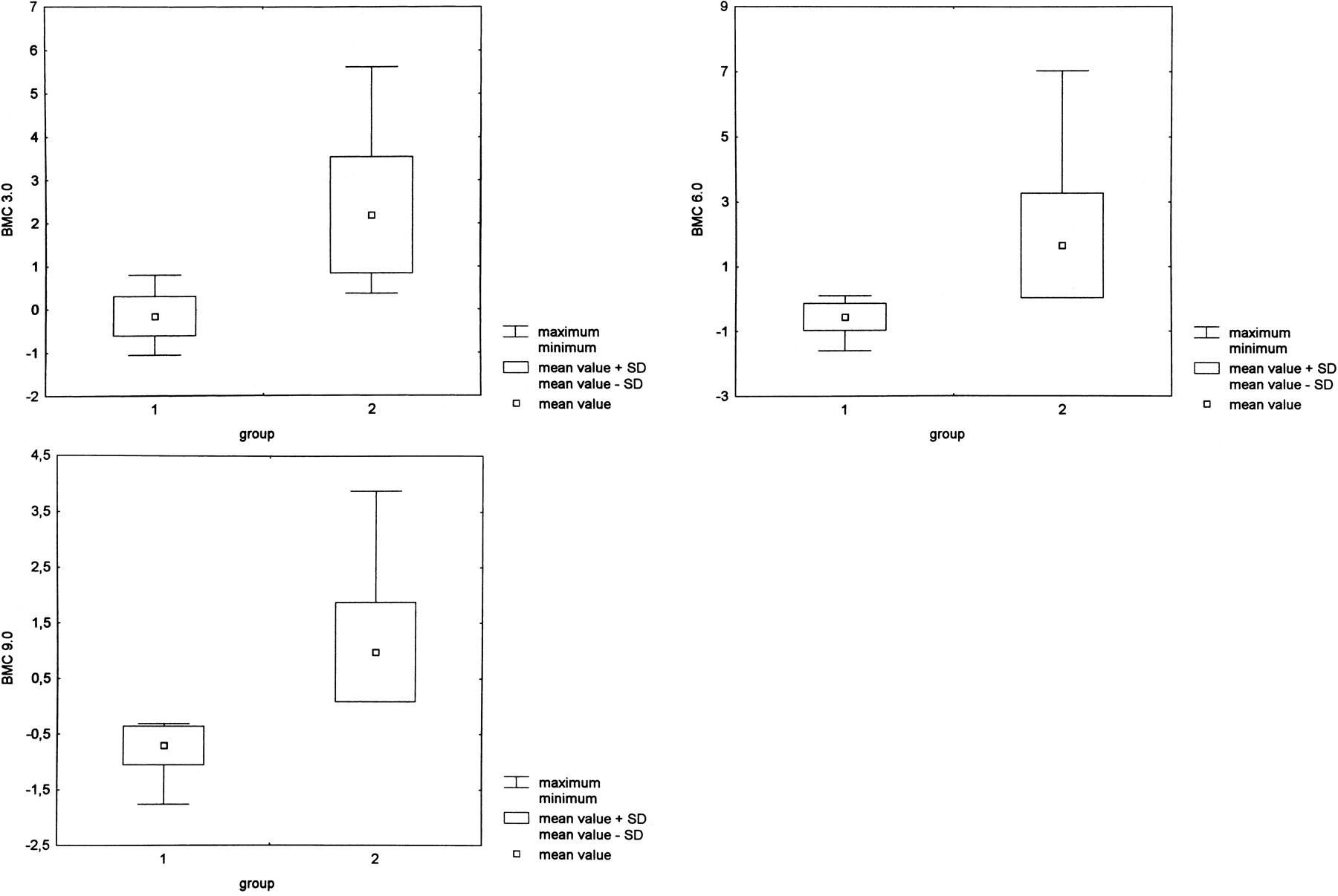
    A–C, At δ = 3 ms, the bone marrow contrast has negative values (mean, −0.16; SD, 0.45) for osteoporotic fractures (group 1) and positive values (mean, 2.19; SD, 1.35) for tumorous fractures (group 2) (A). This represents the hypointense signal in benign versus hyperintense signal in malignant fractures and is statistically different at P < .001. However, there is a small overlap between the maximum values of osteoporotic fractures and the minimum values of malignant fractures at δ = 3 ms. This overlap is reduced with increasing diffusion weighting at δ = 6.0 and 9.0 ms, owing to a signal reduction in benign osteoporotic fractures (B and C).

  • fig 2.
    • Download figure
    • Open in new tab
    • Download powerpoint
    fig 2.

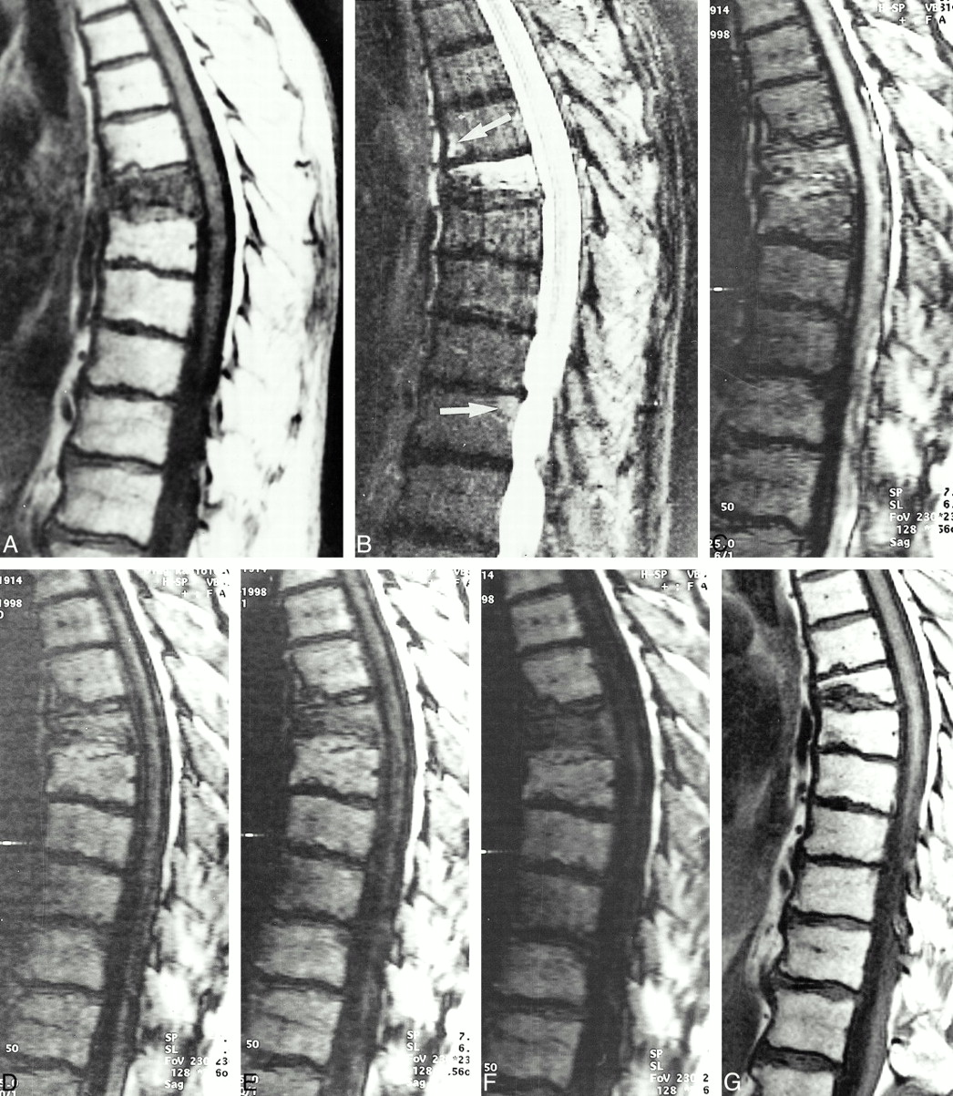
    A, T1-weighted SE image (450/12) in an 84-year-old man with a compression fracture of the seventh thoracic vertebral body. The fracture occurred in the absence of trauma.

    B, Complete replacement of the bone marrow space. STIR image (3600/60, TI = 150) shows hyperintensity in the fractured vertebral body. Note also the focal hyperintensities in the sixth thoracic vertebra (top arrow) anteriorly and in the 11th thoracic vertebra posteriorly and adjacent to the endplate (bottom arrow).

    C, SSFP sequence with very low diffusion weighting (δ = 0.6 ms) shows hyperintense signal in the fractured vertebral body.

    D–F, Increased diffusion weighting with the SSFP sequence (D = 3 ms, E = 6.0 ms, F = 9.0 ms) shows substantial signal loss in the fractured vertebral body. The fractured vertebral body is isointense at δ = 3 ms and markedly hypointense at δ = 6.0 and 9.0 ms. The diagnostic value of increased diffusion weighting is the signal loss in benign bone marrow edema. Note that the focal areas of hyperintensity on the STIR images are also hypointense on the SSFP sequence, indicating benign edema.

    G, Follow-up MR study 8 months later shows healing of the fracture with restitution of fat cells.

  • fig 3.
    • Download figure
    • Open in new tab
    • Download powerpoint
    fig 3.

    62-year-old man with a vertebral fracture due to a metastasis of a transitional cell carcinoma in the fifth thoracic vertebral body.

    A, Hypointensity in the vertebra on T1-weighted SE images.

    B, Corresponding STIR image (3600/60, TI = 150) shows homogeneous high signal intensity in the fractured vertebral body.

    C–F, SSFP image of the same slice with increasing diffusion weighting (C = 0.6 ms, D = 3.0 ms, E = 6.0 ms, and F = 9.0 ms). The signal intensity is markedly hyperintense at δ = 0.6 to 6.0 ms and hyperintense at δ = 9.0 ms. There is a signal loss in the overall image with high diffusion weighting at δ = 9.0 ms. Note the focal area of hyperintensity in the seventh thoracic vertebral body posteriorly (arrow, C). This represents a fat island, because it is hyperintense on T1-weighted SE images and hypointense on STIR images. Fat has an extremely low diffusion coefficient and shows no signal loss on diffusion-weighted images.

Tables

  • Figures
  • Table1

    Qualitative evaluation of signal intensities of fractured vertebral bodies for osteoporotic and tumorous fractures with increasing {δ} relative to normal surrounding bone marrow

PreviousNext
Back to top

In this issue

American Journal of Neuroradiology
Vol. 22, Issue 2
1 Feb 2001
  • Table of Contents
  • Index by author
Advertisement
Print
Download PDF
Email Article

Thank you for your interest in spreading the word on American Journal of Neuroradiology.

NOTE: We only request your email address so that the person you are recommending the page to knows that you wanted them to see it, and that it is not junk mail. We do not capture any email address.

Enter multiple addresses on separate lines or separate them with commas.
Diagnostic Value of Increased Diffusion Weighting of a Steady-state Free Precession Sequence for Differentiating Acute Benign Osteoporotic Fractures from Pathologic Vertebral Compression Fractures
(Your Name) has sent you a message from American Journal of Neuroradiology
(Your Name) thought you would like to see the American Journal of Neuroradiology web site.
CAPTCHA
This question is for testing whether or not you are a human visitor and to prevent automated spam submissions.
Cite this article
Andrea Baur, Armin Huber, Birgit Ertl-Wagner, Roland Dürr, Stefan Zysk, Susanne Arbogast, Michael Deimling, Maximilian Reiser
Diagnostic Value of Increased Diffusion Weighting of a Steady-state Free Precession Sequence for Differentiating Acute Benign Osteoporotic Fractures from Pathologic Vertebral Compression Fractures
American Journal of Neuroradiology Feb 2001, 22 (2) 366-372;

Citation Manager Formats

  • BibTeX
  • Bookends
  • EasyBib
  • EndNote (tagged)
  • EndNote 8 (xml)
  • Medlars
  • Mendeley
  • Papers
  • RefWorks Tagged
  • Ref Manager
  • RIS
  • Zotero
0 Responses
Respond to this article
Share
Bookmark this article
Diagnostic Value of Increased Diffusion Weighting of a Steady-state Free Precession Sequence for Differentiating Acute Benign Osteoporotic Fractures from Pathologic Vertebral Compression Fractures
Andrea Baur, Armin Huber, Birgit Ertl-Wagner, Roland Dürr, Stefan Zysk, Susanne Arbogast, Michael Deimling, Maximilian Reiser
American Journal of Neuroradiology Feb 2001, 22 (2) 366-372;
del.icio.us logo Twitter logo Facebook logo Mendeley logo
  • Tweet Widget
  • Facebook Like
  • Google Plus One

Jump to section

  • Article
    • Abstract
    • Methods
    • Results
    • Discussion
    • Conclusion
    • Footnotes
    • References
  • Figures & Data
  • Info & Metrics
  • Responses
  • References
  • PDF

Related Articles

  • No related articles found.
  • PubMed
  • Google Scholar

Cited By...

  • Review of the Imaging Features of Benign Osteoporotic and Malignant Vertebral Compression Fractures
  • Imaging in myeloma
  • Diffusion-Weighted Imaging of the Spine: Is It Reliable?
  • Crossref
  • Google Scholar

This article has not yet been cited by articles in journals that are participating in Crossref Cited-by Linking.

More in this TOC Section

  • CT-Guided C2 Dorsal Root Ganglion Radiofrequency Ablation for the Treatment of Cervicogenic Headache: Case Series and Clinical Outcomes
  • The Evaluation and Prediction of Laminoplasty Surgery Outcome in Patients with Degenerative Cervical Myelopathy Using Diffusion Tensor MRI
  • Subject-Specific Studies of CSF Bulk Flow Patterns in the Spinal Canal: Implications for the Dispersion of Solute Particles in Intrathecal Drug Delivery
Show more Spine

Similar Articles

Advertisement

Indexed Content

  • Current Issue
  • Accepted Manuscripts
  • Article Preview
  • Past Issues
  • Editorials
  • Editor's Choice
  • Fellows' Journal Club
  • Letters to the Editor
  • Video Articles

Cases

  • Case Collection
  • Archive - Case of the Week
  • Archive - Case of the Month
  • Archive - Classic Case

More from AJNR

  • Trainee Corner
  • Imaging Protocols
  • MRI Safety Corner
  • Book Reviews

Multimedia

  • AJNR Podcasts
  • AJNR Scantastics

Resources

  • Turnaround Time
  • Submit a Manuscript
  • Submit a Video Article
  • Submit an eLetter to the Editor/Response
  • Manuscript Submission Guidelines
  • Statistical Tips
  • Fast Publishing of Accepted Manuscripts
  • Graphical Abstract Preparation
  • Imaging Protocol Submission
  • Evidence-Based Medicine Level Guide
  • Publishing Checklists
  • Author Policies
  • Become a Reviewer/Academy of Reviewers
  • News and Updates

About Us

  • About AJNR
  • Editorial Board
  • Editorial Board Alumni
  • Alerts
  • Permissions
  • Not an AJNR Subscriber? Join Now
  • Advertise with Us
  • Librarian Resources
  • Feedback
  • Terms and Conditions
  • AJNR Editorial Board Alumni

American Society of Neuroradiology

  • Not an ASNR Member? Join Now

© 2025 by the American Society of Neuroradiology All rights, including for text and data mining, AI training, and similar technologies, are reserved.
Print ISSN: 0195-6108 Online ISSN: 1936-959X

Powered by HighWire