Advertisement

Main menu

  • Home
  • Content
    • Current Issue
    • Accepted Manuscripts
    • Article Preview
    • Past Issue Archive
    • Video Articles
    • AJNR Case Collection
    • Case of the Week Archive
    • Case of the Month Archive
    • Classic Case Archive
  • Special Collections
    • AJNR Awards
    • Low-Field MRI
    • Alzheimer Disease
    • ASNR Foundation Special Collection
    • Photon-Counting CT
    • View All
  • Multimedia
    • AJNR Podcasts
    • AJNR SCANtastic
    • Trainee Corner
    • MRI Safety Corner
    • Imaging Protocols
  • For Authors
    • Submit a Manuscript
    • Submit a Video Article
    • Submit an eLetter to the Editor/Response
    • Manuscript Submission Guidelines
    • Statistical Tips
    • Fast Publishing of Accepted Manuscripts
    • Graphical Abstract Preparation
    • Imaging Protocol Submission
    • Author Policies
  • About Us
    • About AJNR
    • Editorial Board
    • Editorial Board Alumni
  • More
    • Become a Reviewer/Academy of Reviewers
    • Subscribers
    • Permissions
    • Alerts
    • Feedback
    • Advertisers
    • ASNR Home

User menu

  • Alerts
  • Log in

Search

  • Advanced search
American Journal of Neuroradiology

American Journal of Neuroradiology

ASHNR American Society of Functional Neuroradiology ASHNR American Society of Pediatric Neuroradiology ASSR
  • Alerts
  • Log in

Advanced Search

  • Home
  • Content
    • Current Issue
    • Accepted Manuscripts
    • Article Preview
    • Past Issue Archive
    • Video Articles
    • AJNR Case Collection
    • Case of the Week Archive
    • Case of the Month Archive
    • Classic Case Archive
  • Special Collections
    • AJNR Awards
    • Low-Field MRI
    • Alzheimer Disease
    • ASNR Foundation Special Collection
    • Photon-Counting CT
    • View All
  • Multimedia
    • AJNR Podcasts
    • AJNR SCANtastic
    • Trainee Corner
    • MRI Safety Corner
    • Imaging Protocols
  • For Authors
    • Submit a Manuscript
    • Submit a Video Article
    • Submit an eLetter to the Editor/Response
    • Manuscript Submission Guidelines
    • Statistical Tips
    • Fast Publishing of Accepted Manuscripts
    • Graphical Abstract Preparation
    • Imaging Protocol Submission
    • Author Policies
  • About Us
    • About AJNR
    • Editorial Board
    • Editorial Board Alumni
  • More
    • Become a Reviewer/Academy of Reviewers
    • Subscribers
    • Permissions
    • Alerts
    • Feedback
    • Advertisers
    • ASNR Home
  • Follow AJNR on Twitter
  • Visit AJNR on Facebook
  • Follow AJNR on Instagram
  • Join AJNR on LinkedIn
  • RSS Feeds

AJNR Awards, New Junior Editors, and more. Read the latest AJNR updates

Research ArticleBrain

Plasticity of the Human Motor Cortex in Patients with Arteriovenous Malformations: A Functional MR Imaging Study

Hatem Alkadhi, Spyros S. Kollias, Gérard R. Crelier, Xavier Golay, Marie-Claude Hepp-Reymond and Anton Valavanis
American Journal of Neuroradiology September 2000, 21 (8) 1423-1433;
Hatem Alkadhi
  • Find this author on Google Scholar
  • Find this author on PubMed
  • Search for this author on this site
Spyros S. Kollias
  • Find this author on Google Scholar
  • Find this author on PubMed
  • Search for this author on this site
Gérard R. Crelier
  • Find this author on Google Scholar
  • Find this author on PubMed
  • Search for this author on this site
Xavier Golay
  • Find this author on Google Scholar
  • Find this author on PubMed
  • Search for this author on this site
Marie-Claude Hepp-Reymond
  • Find this author on Google Scholar
  • Find this author on PubMed
  • Search for this author on this site
Anton Valavanis
  • Find this author on Google Scholar
  • Find this author on PubMed
  • Search for this author on this site
  • Article
  • Figures & Data
  • Info & Metrics
  • Responses
  • References
  • PDF
Loading

Article Figures & Data

Figures

  • Tables
  • fig 1.
    • Download figure
    • Open in new tab
    • Download powerpoint
    fig 1.

    Patient 1.

    A, Sagittal T1-weighted image (500/14/4) shows the AVM in the left precentral gyrus and central sulcus, thereby involving the expected primary representation area of the left hand.

    B and C, Four adjacent axial sections show cortical maps related to finger movements of the left (B) and right (C) hand. Compared with the right hemisphere M1 representation, which is in the expected anatomic location (B), activation in the left hemisphere is displaced laterally. This displacement does not follow the structural distortion of the rolandic region induced by the AVM. Note further activation in bilateral parietal and SMAs.

    fig A1. Plotted signal models of the boxcar functions with different time delays (one, two, and three TRs; upper row), and the trapezoidal function with a one-TR delay and a rise time of two TRs (lower image)

  • fig 2.
    • Download figure
    • Open in new tab
    • Download powerpoint
    fig 2.

    Patient 3.

    A, Sagittal T1-weighted image (500/14/4) shows an AVM in the right precentral gyrus, involving the anatomically expected area of hand representation.

    B and C, As compared with the unaffected left hemisphere (B), the hand representation in the affected right hemisphere (C) is displaced medially within the precentral gyrus. Additional activation within the ipsilateral M1 is detected. The functional displacement does not follow the structural distortion induced by the underlying disorder. Note additional activation in bilateral supplementary motor (arrows, upper right image in C) and parietal areas.

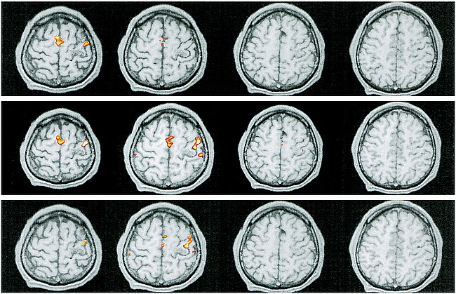
    fig A2. Patient 1. Reprocessed functional MR imaging experiment using a boxcar function with delays of one TR (upper row), two TRs (middle row), and three TRs (lower row)

  • fig 3.
    • Download figure
    • Open in new tab
    • Download powerpoint
    fig 3.

    Patient 6.

    A, Sagittal T1-weighted image (500/14/4) shows the relationship between the left-sided, central AVM and the precentral knob.

    B, Movements of the right foot show the expected response in the left paracentral lobule (contralateral M1) with additional activation in bilateral SMAs.

    C, Right-hand movements do not elicit activation in M1 but there is prominent signal in bilateral SMAs. Note additional activation in ipsilateral dorsal premotor areas and in the contralateral parietal areas and CMA.

    fig A3. Patient 6. Reprocessed functional MR imaging experiment using a boxcar function with delays of one TR (upper row), two TRs (middle row), and three TRs (lower row)

  • fig 4.
    • Download figure
    • Open in new tab
    • Download powerpoint
    fig 4.

    Patient 7.

    A, T1-weighted sagittal image (500/14/4) shows an extensive AVM in the right paracentral lobule extending to the precuneus, thereby involving the expected primary representation of the left foot.

    B, Left-hand movements show the expected activation in the contralateral M1.

    C, Flexion and extension of the left foot reveal absent contralateral activation but prominent activation in the ipsilateral M1, SMA, dorsal premotor area, and CMA, and in the parietal areas bilaterally.

    fig A4. Reprocessed functional MR imaging experiment using a trapezoidal function with a delay of one TR and a rise time of two TRs in patients 1 (upper row) and 6 (lower row)

Tables

  • Figures
  • TABLE 1:
    • View popup
    • Download powerpoint
    TABLE 1:

    Characteristics of patients and arteriovenous malformations (AVMs)

  • TABLE 2:
    • View popup
    • Download powerpoint
    TABLE 2:

    Activated primary and nonprimary motor areas during contralesional limb movements*

PreviousNext
Back to top

In this issue

American Journal of Neuroradiology
Vol. 21, Issue 8
1 Sep 2000
  • Table of Contents
  • Index by author
Advertisement
Print
Download PDF
Email Article

Thank you for your interest in spreading the word on American Journal of Neuroradiology.

NOTE: We only request your email address so that the person you are recommending the page to knows that you wanted them to see it, and that it is not junk mail. We do not capture any email address.

Enter multiple addresses on separate lines or separate them with commas.
Plasticity of the Human Motor Cortex in Patients with Arteriovenous Malformations: A Functional MR Imaging Study
(Your Name) has sent you a message from American Journal of Neuroradiology
(Your Name) thought you would like to see the American Journal of Neuroradiology web site.
CAPTCHA
This question is for testing whether or not you are a human visitor and to prevent automated spam submissions.
Cite this article
Hatem Alkadhi, Spyros S. Kollias, Gérard R. Crelier, Xavier Golay, Marie-Claude Hepp-Reymond, Anton Valavanis
Plasticity of the Human Motor Cortex in Patients with Arteriovenous Malformations: A Functional MR Imaging Study
American Journal of Neuroradiology Sep 2000, 21 (8) 1423-1433;

Citation Manager Formats

  • BibTeX
  • Bookends
  • EasyBib
  • EndNote (tagged)
  • EndNote 8 (xml)
  • Medlars
  • Mendeley
  • Papers
  • RefWorks Tagged
  • Ref Manager
  • RIS
  • Zotero
0 Responses
Respond to this article
Share
Bookmark this article
Plasticity of the Human Motor Cortex in Patients with Arteriovenous Malformations: A Functional MR Imaging Study
Hatem Alkadhi, Spyros S. Kollias, Gérard R. Crelier, Xavier Golay, Marie-Claude Hepp-Reymond, Anton Valavanis
American Journal of Neuroradiology Sep 2000, 21 (8) 1423-1433;
del.icio.us logo Twitter logo Facebook logo Mendeley logo
  • Tweet Widget
  • Facebook Like
  • Google Plus One

Jump to section

  • Article
    • Abstract
    • Methods
    • Results
    • Discussion
    • Conclusion
    • Footnotes
    • References
  • Figures & Data
  • Info & Metrics
  • Responses
  • References
  • PDF

Related Articles

  • No related articles found.
  • PubMed
  • Google Scholar

Cited By...

  • Classification of brain arteriovenous malformations located in motor-related areas based on location and anterior choroidal artery feeding
  • Preoperative motor system brain mapping using positron emission tomography and statistical parametric mapping: hints on cortical reorganisation
  • Activation in primary and secondary motor areas in patients with CNS neoplasms and weakness
  • Metabolic and electrophysiological validation of functional MRI
  • Crossref
  • Google Scholar

This article has not yet been cited by articles in journals that are participating in Crossref Cited-by Linking.

More in this TOC Section

  • Optimal MRI Sequence for Identifying Occlusion Location in Acute Stroke: Which Value of Time-Resolved Contrast-Enhanced MRA?
  • Evaluating the Effects of White Matter Multiple Sclerosis Lesions on the Volume Estimation of 6 Brain Tissue Segmentation Methods
  • Quiet PROPELLER MRI Techniques Match the Quality of Conventional PROPELLER Brain Imaging Techniques
Show more Brain

Similar Articles

Advertisement

Indexed Content

  • Current Issue
  • Accepted Manuscripts
  • Article Preview
  • Past Issues
  • Editorials
  • Editor's Choice
  • Fellows' Journal Club
  • Letters to the Editor
  • Video Articles

Cases

  • Case Collection
  • Archive - Case of the Week
  • Archive - Case of the Month
  • Archive - Classic Case

More from AJNR

  • Trainee Corner
  • Imaging Protocols
  • MRI Safety Corner
  • Book Reviews

Multimedia

  • AJNR Podcasts
  • AJNR Scantastics

Resources

  • Turnaround Time
  • Submit a Manuscript
  • Submit a Video Article
  • Submit an eLetter to the Editor/Response
  • Manuscript Submission Guidelines
  • Statistical Tips
  • Fast Publishing of Accepted Manuscripts
  • Graphical Abstract Preparation
  • Imaging Protocol Submission
  • Evidence-Based Medicine Level Guide
  • Publishing Checklists
  • Author Policies
  • Become a Reviewer/Academy of Reviewers
  • News and Updates

About Us

  • About AJNR
  • Editorial Board
  • Editorial Board Alumni
  • Alerts
  • Permissions
  • Not an AJNR Subscriber? Join Now
  • Advertise with Us
  • Librarian Resources
  • Feedback
  • Terms and Conditions
  • AJNR Editorial Board Alumni

American Society of Neuroradiology

  • Not an ASNR Member? Join Now

© 2025 by the American Society of Neuroradiology All rights, including for text and data mining, AI training, and similar technologies, are reserved.
Print ISSN: 0195-6108 Online ISSN: 1936-959X

Powered by HighWire