
Article Figures & Data
Figures
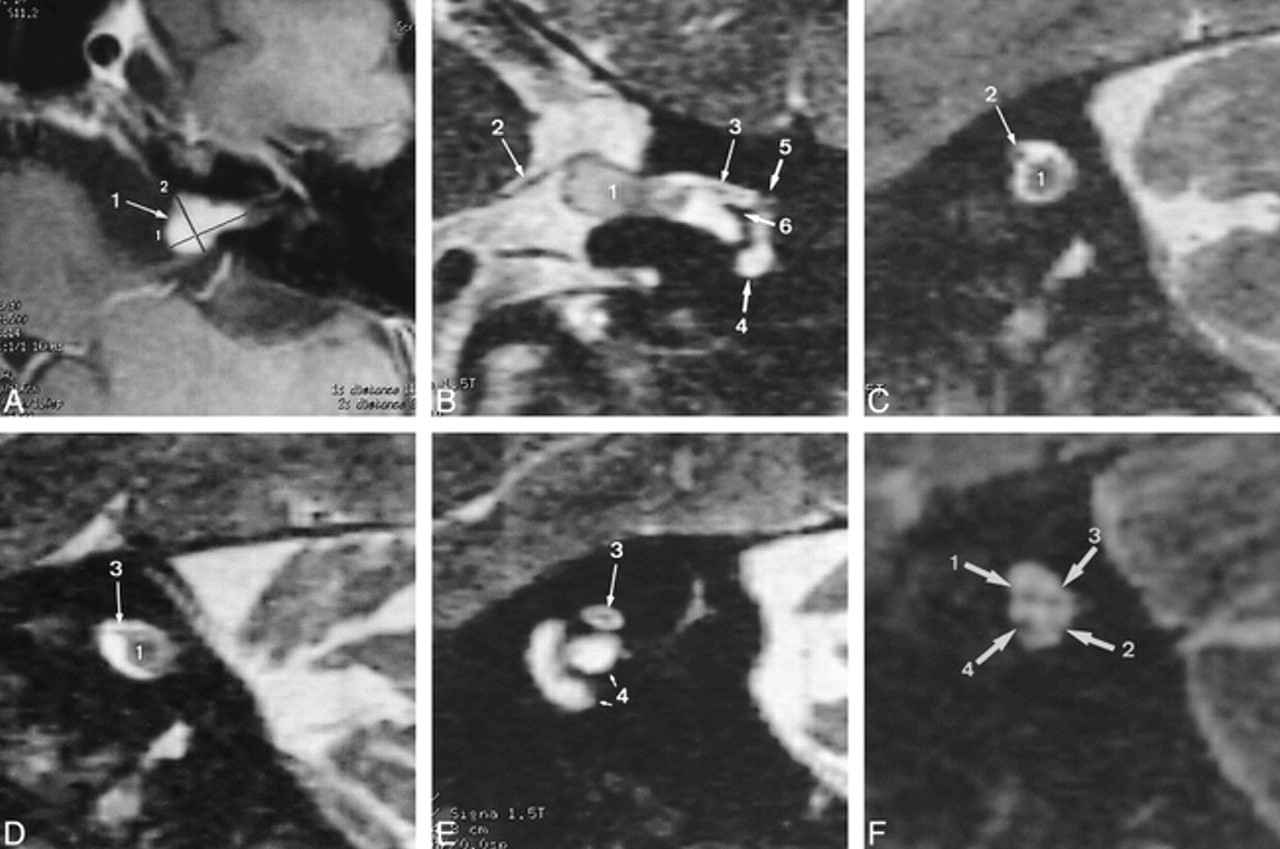
- FIG 1.
A, Postcontrast T1-weighted transverse MR image (600/16/4) shows a vestibular schwannoma (large 1), 11 × 8 × 5 mm in size, with both intra- and extrameatal tumor portions (crosshatches 1 and 2 indicate the measured tumor diameters).
B, Reformatted oblique coronal 3D T2-weighted FSE image (4000/150/1) shows the facial nerve (2, 3) in an anterosuperior position within the IAC in relation to the tumor (1). The medial (2) and lateral (3) segments are delineated. The nerve has a normal diameter of 1 mm along the medial segment and the medial part of the intervening segment (2), and is focally thinned along the lateral segment and the lateral part of the intervening segment (3). The signal intensity of the thinned lateral segment (3) is slightly increased relative to the medial segment (2) and to the contralateral lateral facial nerve segment (see fig 1F). The cochlea (4), meatal foramen (5), and crista falciformis (6) are depicted.
C–E, Reformatted oblique sagittal images from medial to lateral.
C, Reformatted oblique sagittal 3D T2-weighted FSE image (4000/150/1) along the medial part of the intervening segment. The facial nerve (2) is located anterosuperiorly relative to the tumor (1).
D, Reformatted oblique sagittal 3D T2-weighted FSE image (4000/150/1) along the lateral part of the intervening segment. The facial nerve (3) is still in an anterosuperior position relative to the tumor (1) and is focally thinned with slightly increased signal intensity as compared with the medial segment or with the contralateral facial nerve segment (see fig 1F).
E, Reformatted oblique sagittal 3D T2-weighted FSE image (4000/150/1) along the lateral nerve segment (3) shows a focally thinned lateral nerve segment with slightly increased signal intensity at the entrance of the nerve into the meatal foramen. 4 indicates the cochlea.
F, For comparison, reformatted oblique sagittal 3D T2-weighted FSE image (4000/150/1) through the IAC shows the normal contralateral facial nerve (1) as well as the inferior (2) and superior (3) vestibular nerve and the cochlear nerve (4).
- FIG 2.
A, Postcontrast transverse T1-weighted SE image (600/16/4) shows the intra- and extrameatal location of a vestibular schwannoma (size, 12 × 10 × 7 mm).
B, Reformatted oblique coronal 3D T2-weighted FSE image (4000/150/1) shows the vestibular schwannoma (1) and the lateral part of the intervening segment as well as the lateral segment of the facial nerve (2). The nerve is slightly hyperintense relative to the contralateral lateral facial nerve segment (not shown). Cochlea (4), meatal foramen (5), and crista falciformis (6) are depicted.
C, Reformatted oblique sagittal 3D T2-weighted FSE image (4000/150/1) along the intervening facial nerve segment (2) shows the vestibular schwannoma (1) and the facial nerve shifted to a posterosuperior position. The vestibular nerve is not identified.
D, Reformatted oblique sagittal 3D T2-weighted FSE image (4000/150/1) along the intervening facial nerve segment (2), lateral to C, again shows the vestibular schwannoma (1) and the facial nerve (2) shifted to a posterosuperior position. The vestibular nerve (3) may be assumed to be in a posteroinferior position.
E, Reformatted oblique sagittal 3D T2-weighted FSE image (4000/150/1) along the lateral facial nerve segment (2) shows the facial nerve proximal to its entrance into the bony canal at the meatal foramen. Cochlea (4) and semicircular canals (5) are depicted.
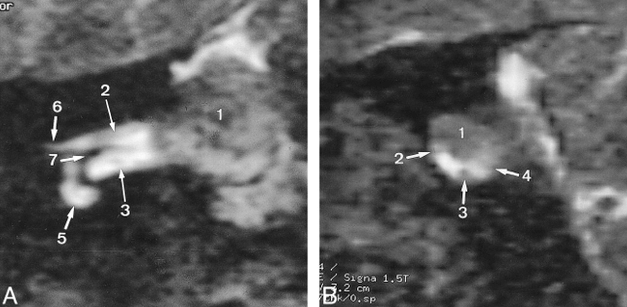
- FIG 3.
A, Reformatted oblique sagittal 3D T2-weighted FSE image (4000/150/1) medial to the tumor shows the superior vestibular nerve (2), the facial nerve (3), the cochlear nerve (4), and the inferior vestibular nerve (5). The facial nerve has a normal diameter and does not show increased signal intensity.
B, Reformatted oblique sagittal 3D T2-weighted FSE image (4000/150/1) along the intervening facial nerve segment shows a small vestibular schwannoma (1) (mean diameter, 3 mm). The tumor most probably arises from the superior vestibular nerve. The facial nerve along its intervening segment (3) is depicted in an anterosuperior position. The cochlear nerve (4) and the inferior vestibular nerve (5) are depicted.
C, Reformatted oblique sagittal 3D T2-weighted FSE image (4000/150/1) lateral to the tumor shows the lateral segment of the facial nerve (3) just proximal to the entrance of the nerve into the bony canal at the meatal foramen. Cochlea (6) and semicircular canal (7) are depicted.
D, Reformatted oblique coronal 3D T2-weighted FSE image (4000/150/1) shows the facial nerve (3) along its medial segment as well as along the intervening segment adjacent to the vestibular schwannoma (1). The nerve is in an anterosuperior position.
- FIG 4.
A, Reformatted oblique coronal 3D T2-weighted FSE image (4000/150/1) shows the intra- and extrameatal location of a vestibular schwannoma (1) (size, 13 × 18 × 11 mm). The lateral segment of the facial nerve (2) and the cochlear nerve (3) are depicted, as are the cochlea (5), meatal foramen (6), and crista falciformis (7).
B, Reformatted oblique sagittal 3D T2-weighted FSE image (4000/150/1) shows the vestibular schwannoma (1) and the facial nerve along its intervening segment (2). Because of its position, the tumor (1) most probably arises from the superior vestibular nerve; the facial nerve is shifted to an anteroinferior position. The cochlear nerve (3) and the inferior vestibular nerve (4) are also depicted.


Tables
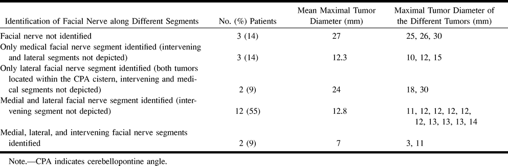
 Table 1:
Identification of different facial nerve segments (medial, lateral, and intervening) adjacent to a vestibular schwannoma
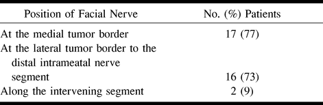
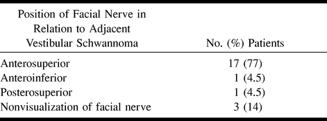
- Table 3:
Spatial relationship between facial nerve and adjacent vestibular schwannoma


