
Article Figures & Data
Figures
- fig 1.
Case 1: 33-year-old woman with sudden onset of severe headache and vomiting.
A, Right visual field at time of presentation shows a right inferior nasal field defect that is complete (dark black region).
B and C, Right visual field 3 weeks (B) and 6 weeks (C) later shows spontaneous progressive improvement of the right inferonasal field defect.
D and E, Noncontrast (D) and contrast-enhanced (E) axial T1-weighted SE images (450/20/2) show an enhancing inflammatory mass surrounding the previously coiled aneurysm. Note the adjacent right optic nerve (arrow, E).
F, Axial FLAIR image (8000/120/2, TI = 2200) shows increased signal intensity, consistent with edema, in the adjacent brain parenchyma.
G, Coronal T1-weighted SE image with fat suppression after IV administration of contrast material shows marked enhancement adjacent to the right optic nerve (arrow). Apparent hyperintensity of both optic nerves represents artifact related to technique, not enhancement, as other images showed no enhancement.
H, Axial T2-weighted GRASE image (5000/100/3) shows the mass to be relatively uniformly and solidly hypointense rather than predominantly hyperintense with a hypointense rim, as would be expected with an abscess.
- fig 2.
Case 2: 63-year-old woman with 7- to 8-mm left supraclinoid ICA aneurysm near origin of ophthalmic artery.
A, Left visual field at time of presentation shows a large nasal scotoma.
B, Left visual field approximately 2 months later (after 7 weeks of corticosteroid treatment) shows significant improvement, with a smaller nasal defect and a paracentral scotoma.
C–E, Contiguous contrast-enhanced axial T1-weighted SE (450/13/2) images show a flow void within the aneurysm (arrow, C) and a surrounding enhancing inflammatory mass that abuts the left optic nerve (arrowheads, D), chiasm (arrow, E), and tract. Note the incidental finding of a developmental venous anomaly in the right temporal lobe.
F, Axial FLAIR image (7000/130, TI = 2200) shows high signal intensity, consistent with edema, in the optic tracts, left greater than right (arrows). Increased signal intensity in the right subinsular region represents chronic microvascular ischemic changes, unrelated to the muslinoma.
G and H, Follow-up MR images 7 weeks later, after corticosteroid treatment. Contrast-enhanced axial T1-weighted SE image (G) shows a persistent enhancing mass, unchanged in size and still involving the left optic pathways. Axial FLAIR image (H) shows mild improvement in edema in the left optic tract.


Tables
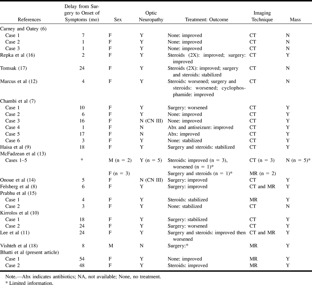
 Table 1:
Muslinoma: summary characteristics of visual and nonvisual deficits
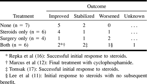
- Table 2:
Summary of final treatment outcome of muslin-induced visual loss (23 cases)


