Advertisement

Main menu

  • Home
  • Content
    • Current Issue
    • Accepted Manuscripts
    • Article Preview
    • Past Issue Archive
    • Video Articles
    • AJNR Case Collection
    • Case of the Week Archive
    • Case of the Month Archive
    • Classic Case Archive
  • Special Collections
    • AJNR Awards
    • Low-Field MRI
    • Alzheimer Disease
    • ASNR Foundation Special Collection
    • Photon-Counting CT
    • View All
  • Multimedia
    • AJNR Podcasts
    • AJNR SCANtastic
    • Trainee Corner
    • MRI Safety Corner
    • Imaging Protocols
  • For Authors
    • Submit a Manuscript
    • Submit a Video Article
    • Submit an eLetter to the Editor/Response
    • Manuscript Submission Guidelines
    • Statistical Tips
    • Fast Publishing of Accepted Manuscripts
    • Graphical Abstract Preparation
    • Imaging Protocol Submission
    • Author Policies
  • About Us
    • About AJNR
    • Editorial Board
    • Editorial Board Alumni
  • More
    • Become a Reviewer/Academy of Reviewers
    • Subscribers
    • Permissions
    • Alerts
    • Feedback
    • Advertisers
    • ASNR Home

User menu

  • Alerts
  • Log in

Search

  • Advanced search
American Journal of Neuroradiology

American Journal of Neuroradiology

ASHNR American Society of Functional Neuroradiology ASHNR American Society of Pediatric Neuroradiology ASSR
  • Alerts
  • Log in

Advanced Search

  • Home
  • Content
    • Current Issue
    • Accepted Manuscripts
    • Article Preview
    • Past Issue Archive
    • Video Articles
    • AJNR Case Collection
    • Case of the Week Archive
    • Case of the Month Archive
    • Classic Case Archive
  • Special Collections
    • AJNR Awards
    • Low-Field MRI
    • Alzheimer Disease
    • ASNR Foundation Special Collection
    • Photon-Counting CT
    • View All
  • Multimedia
    • AJNR Podcasts
    • AJNR SCANtastic
    • Trainee Corner
    • MRI Safety Corner
    • Imaging Protocols
  • For Authors
    • Submit a Manuscript
    • Submit a Video Article
    • Submit an eLetter to the Editor/Response
    • Manuscript Submission Guidelines
    • Statistical Tips
    • Fast Publishing of Accepted Manuscripts
    • Graphical Abstract Preparation
    • Imaging Protocol Submission
    • Author Policies
  • About Us
    • About AJNR
    • Editorial Board
    • Editorial Board Alumni
  • More
    • Become a Reviewer/Academy of Reviewers
    • Subscribers
    • Permissions
    • Alerts
    • Feedback
    • Advertisers
    • ASNR Home
  • Follow AJNR on Twitter
  • Visit AJNR on Facebook
  • Follow AJNR on Instagram
  • Join AJNR on LinkedIn
  • RSS Feeds

AJNR Awards, New Junior Editors, and more. Read the latest AJNR updates

Getting new auth cookie, if you see this message a lot, tell someone!
OtherBrain

Corpus Callosum Infarcts with Atypical Clinical and Radiologic Presentations

David L. Kasow, Sylvie Destian, Carl Braun, John C. Quintas, Nolan J. Kagetsu and Carl E. Johnson
American Journal of Neuroradiology November 2000, 21 (10) 1876-1880;
David L. Kasow
  • Find this author on Google Scholar
  • Find this author on PubMed
  • Search for this author on this site
Sylvie Destian
  • Find this author on Google Scholar
  • Find this author on PubMed
  • Search for this author on this site
Carl Braun
  • Find this author on Google Scholar
  • Find this author on PubMed
  • Search for this author on this site
John C. Quintas
  • Find this author on Google Scholar
  • Find this author on PubMed
  • Search for this author on this site
Nolan J. Kagetsu
  • Find this author on Google Scholar
  • Find this author on PubMed
  • Search for this author on this site
Carl E. Johnson
  • Find this author on Google Scholar
  • Find this author on PubMed
  • Search for this author on this site
  • Article
  • Figures & Data
  • Info & Metrics
  • Responses
  • References
  • PDF
Loading

Article Figures & Data

Figures

  • fig 1.
    • Download figure
    • Open in new tab
    • Download powerpoint
    fig 1.

    Case 1. Axial postcontrast CT image (A) and coronal postcontrast T1-weighted image (B) (480/20/1 [TR/TE/excitations]) show a non-enhancing lesion within both sides of the genu (curved arrow) of the corpus callosum with mass effect on the frontal horns. Axial T2-weighted image (C) (2500/105/1) shows corresponding high signal intensity within the genu of the corpus callosum (curved arrow), crossing the midline. An old infarct is noted within the right posterior border zone and scattered foci of T2 signal abnormality are seen in the left frontal and parietal regions. A biopsy of the lesion was obtained, confirming an infarct

  • fig 2.
    • Download figure
    • Open in new tab
    • Download powerpoint
    fig 2.

    Case 2. Postcontrast axial T1-weighted image (A) (500/20/1) shows subtle enhancement with surrounding hypointensity within the genu of the corpus callosum (arrows). FLAIR images (B, C) (8900/140/1) show abnormal increased signal within both sides of the genu and body of the corpus callosum. Axial FLAIR images (D, E) (8900/140/1) from follow-up MR imaging performed 3 weeks later showed almost complete resolution of the abnormal signal within the corpus callosum

  • fig 3.
    • Download figure
    • Open in new tab
    • Download powerpoint
    fig 3.

    Case 3. Sagittal precontrast T1-weighted image (A) (400/12/2) shows a hypointense ovoid mass (arrow) within the body of the corpus callosum, depressing the roof of the lateral ventricle. After administration of contrast material (B) (400/12/2), it enhances homogeneously. A small infarct (white arrow) is noted within the right thalamus on the axial T2-weighted image (C) (2560/90/1). Subsequent biopsy confirmed an infarct
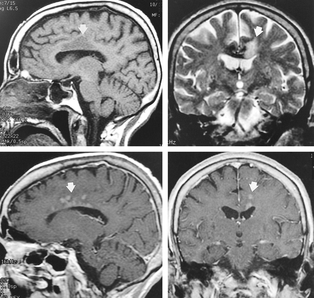
  • fig 4.
    • Download figure
    • Open in new tab
    • Download powerpoint
    fig 4.

    Case 4. Sagittal non-contrast T1- (A) (400/10/1) and coronal T2-weighted (B) (2500/105/1) images show abnormal signal within the body of the corpus callosum and adjacent white matter (arrow) on the left side. Sagittal (C) (400/8/2) and coronal (D) (450/20/1) postcontrast T1-weighted images from a subsequent MR examination show abnormal enhancement (arrow) within the corpus callosum and adjacent white matter

PreviousNext
Back to top

In this issue

American Journal of Neuroradiology
Vol. 21, Issue 10
1 Nov 2000
  • Table of Contents
  • Index by author
Advertisement
Print
Download PDF
Email Article

Thank you for your interest in spreading the word on American Journal of Neuroradiology.

NOTE: We only request your email address so that the person you are recommending the page to knows that you wanted them to see it, and that it is not junk mail. We do not capture any email address.

Enter multiple addresses on separate lines or separate them with commas.
Corpus Callosum Infarcts with Atypical Clinical and Radiologic Presentations
(Your Name) has sent you a message from American Journal of Neuroradiology
(Your Name) thought you would like to see the American Journal of Neuroradiology web site.
CAPTCHA
This question is for testing whether or not you are a human visitor and to prevent automated spam submissions.
Cite this article
David L. Kasow, Sylvie Destian, Carl Braun, John C. Quintas, Nolan J. Kagetsu, Carl E. Johnson
Corpus Callosum Infarcts with Atypical Clinical and Radiologic Presentations
American Journal of Neuroradiology Nov 2000, 21 (10) 1876-1880;

Citation Manager Formats

  • BibTeX
  • Bookends
  • EasyBib
  • EndNote (tagged)
  • EndNote 8 (xml)
  • Medlars
  • Mendeley
  • Papers
  • RefWorks Tagged
  • Ref Manager
  • RIS
  • Zotero
0 Responses
Respond to this article
Share
Bookmark this article
Corpus Callosum Infarcts with Atypical Clinical and Radiologic Presentations
David L. Kasow, Sylvie Destian, Carl Braun, John C. Quintas, Nolan J. Kagetsu, Carl E. Johnson
American Journal of Neuroradiology Nov 2000, 21 (10) 1876-1880;
del.icio.us logo Twitter logo Facebook logo Mendeley logo
  • Tweet Widget
  • Facebook Like
  • Google Plus One

Jump to section

  • Article
    • Abstract
    • Case Histories
    • Discussion
    • Conclusion
    • References
  • Figures & Data
  • Info & Metrics
  • Responses
  • References
  • PDF

Related Articles

  • No related articles found.
  • PubMed
  • Google Scholar

Cited By...

  • Rates of local procedural-related structural injury following clipping or coiling of anterior communicating artery aneurysms
  • The corpus callosum: white matter or terra incognita
  • Crossref
  • Google Scholar

This article has not yet been cited by articles in journals that are participating in Crossref Cited-by Linking.

More in this TOC Section

  • Optimal MRI Sequence for Identifying Occlusion Location in Acute Stroke: Which Value of Time-Resolved Contrast-Enhanced MRA?
  • Evaluating the Effects of White Matter Multiple Sclerosis Lesions on the Volume Estimation of 6 Brain Tissue Segmentation Methods
  • Quiet PROPELLER MRI Techniques Match the Quality of Conventional PROPELLER Brain Imaging Techniques
Show more Brain

Similar Articles

Advertisement

Indexed Content

  • Current Issue
  • Accepted Manuscripts
  • Article Preview
  • Past Issues
  • Editorials
  • Editor's Choice
  • Fellows' Journal Club
  • Letters to the Editor
  • Video Articles

Cases

  • Case Collection
  • Archive - Case of the Week
  • Archive - Case of the Month
  • Archive - Classic Case

More from AJNR

  • Trainee Corner
  • Imaging Protocols
  • MRI Safety Corner
  • Book Reviews

Multimedia

  • AJNR Podcasts
  • AJNR Scantastics

Resources

  • Turnaround Time
  • Submit a Manuscript
  • Submit a Video Article
  • Submit an eLetter to the Editor/Response
  • Manuscript Submission Guidelines
  • Statistical Tips
  • Fast Publishing of Accepted Manuscripts
  • Graphical Abstract Preparation
  • Imaging Protocol Submission
  • Evidence-Based Medicine Level Guide
  • Publishing Checklists
  • Author Policies
  • Become a Reviewer/Academy of Reviewers
  • News and Updates

About Us

  • About AJNR
  • Editorial Board
  • Editorial Board Alumni
  • Alerts
  • Permissions
  • Not an AJNR Subscriber? Join Now
  • Advertise with Us
  • Librarian Resources
  • Feedback
  • Terms and Conditions
  • AJNR Editorial Board Alumni

American Society of Neuroradiology

  • Not an ASNR Member? Join Now

© 2025 by the American Society of Neuroradiology All rights, including for text and data mining, AI training, and similar technologies, are reserved.
Print ISSN: 0195-6108 Online ISSN: 1936-959X

Powered by HighWire